Combined ultrasound and endoscopy system

A technology of ultrasound and ultrasound probe, applied in the field of ultrasound and endoscope combined system

Pending Publication Date: 2021-07-23
SUZHOU ACUVU MEDICAL TECH CO LTD
View PDF1 Cites 2 Cited by
  • Summary
  • Abstract
  • Description
  • Claims
  • Application Information

AI Technical Summary

Problems solved by technology

Although endoscopy can be performed before sonohysterography to obtain a direct visual image of the uterine wall, it is usually performed "blindly" or without any real-time visual aid during the insertion of the ultrasound probe Acoustic contrast

Method used


Image

  • Combined ultrasound and endoscopy system
  • Combined ultrasound and endoscopy system
  • Combined ultrasound and endoscopy system

Examples


Embodiment Construction

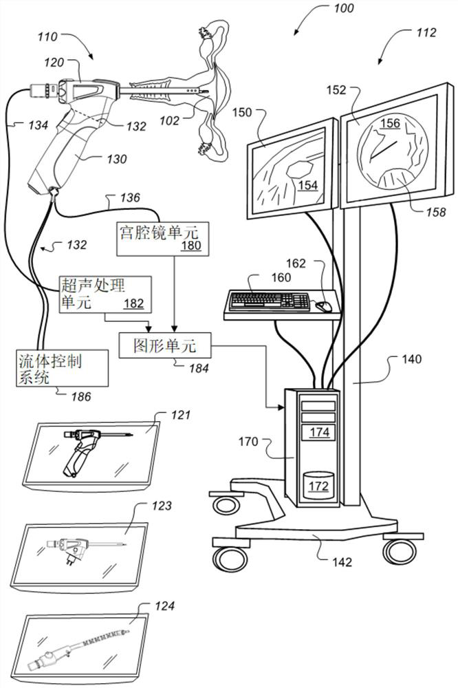
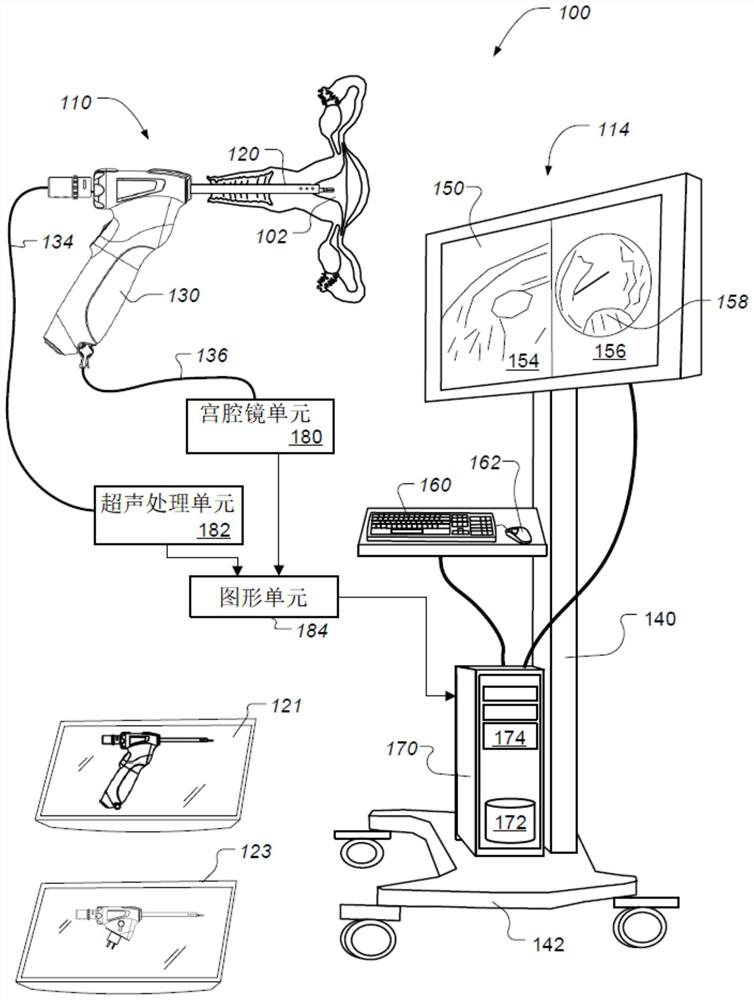
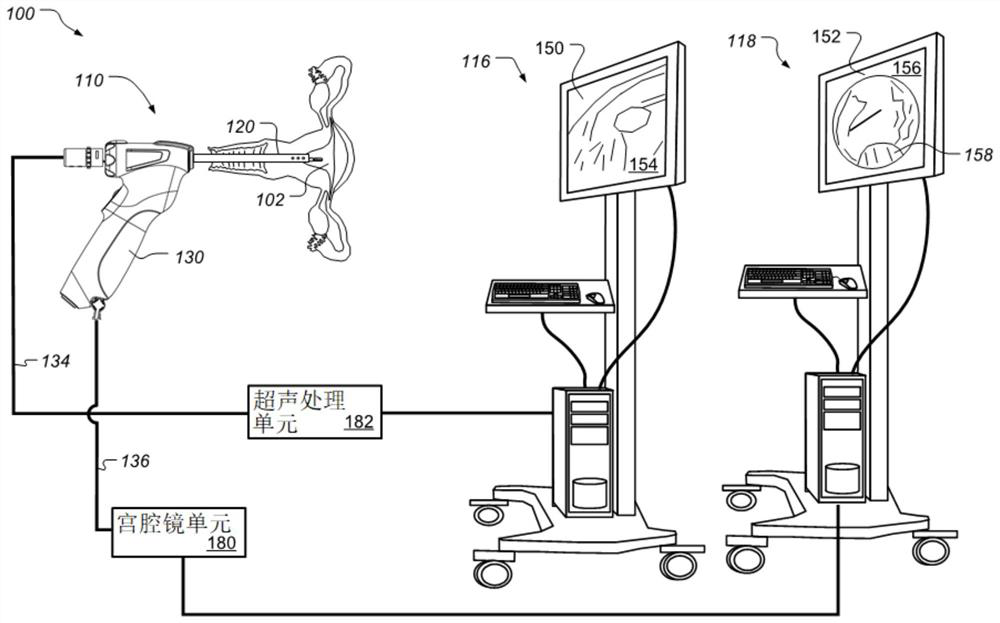
[0025] A detailed description of examples of preferred embodiments is provided below. While several embodiments have been described, it should be understood that the novel subject matter described in this patent specification is not limited to any one or combination of embodiments described herein, but encompasses numerous alternatives, modifications and equivalents. Additionally, although numerous specific details are set forth in the following description in order to provide a thorough understanding, some embodiments may be practiced without some or all of these details. Furthermore, for the sake of clarity, certain technical material that is known in the prior art has not been described in detail to avoid unnecessarily obscuring novel subject matter described herein. It should be clear that various features of one or more specific embodiments described herein may be used in combination with features of other described embodiments or other features. In addition, the same re...


PUM


Abstract

The present invention relates to a combined ultrasound and endoscopy system. The combined ultrasonic and endoscopy system includes a cannulawith a distal tip configured for insertion into an internal organ or other internal body structure. The distal tip is configured to include both an ultrasound probe head and a camera module. The ultrasonic and direct vision endoscopy images can be simultaneously displayed to a user on a display monitor. The ultrasound probe head can be rotated and steered to scan any location in the human organ cavity. The ultrasound probe can be re-usable or single use. The endoscopy system can be configured with a handheld portion that includes a re-usable handle portion and a single use portion that is configured to be disposed of following a single use. The system can also be configured using a conventional re-usable endoscope with working channels and an endoscopy processing tower system.

Description

[0001] Cross References to Related Applications [0002] This application claims priority to U.S. Provisional Application No. 62 / 933,216, filed November 8, 2019, and PCT Application No. PCT / IB2020 / 000470, filed June 11, 2020, which was adopted by Incorporated herein by reference. technical field [0003] This patent specification generally relates to a medical device for examination of tissues and organs. More specifically, some embodiments relate to a combined ultrasound and endoscope system for examining internal organs and other internal body structures. Background technique [0004] Ultrasound is a sound wave with a frequency higher than that audible to humans (>20,000Hz). Ultrasound images, also called sonograms, are made by using a probe to send ultrasound pulses into tissue. Ultrasound pulses echo from tissue with different reflective properties and are recorded and displayed as an image. Typically medical ultrasound is used to create images of internal organs...

Claims


Application Information

Patent Timeline
Patent Type & Authority Applications(China)
IPC IPC(8): A61B1/303A61B1/307A61B8/12A61B1/018A61B1/04A61B1/06
CPCA61B1/303A61B1/307A61B8/12A61B1/04A61B1/00009A61B1/018A61B1/0676A61B1/015A61B8/445A61B8/463A61B1/000094
Inventor 俞增平
Owner SUZHOU ACUVU MEDICAL TECH CO LTD
Why Eureka
Try Eureka