Interventional contrast imaging device and method

An angiographic imaging and support plate technology, applied in the field of medical devices, can solve the problems of inconvenience in carrying patients and changing the bed back and forth, and achieve the effects of convenient closing, easy opening and reasonable design

Pending Publication Date: 2021-08-20
XIEHE HOSPITAL ATTACHED TO TONGJI MEDICAL COLLEGE HUAZHONG SCI & TECH UNIV
View PDF13 Cites 0 Cited by
  • Summary
  • Abstract
  • Description
  • Claims
  • Application Information

AI Technical Summary

Problems solved by technology

[0004] The present invention proposes an interventional contrast imaging device and method, which solves the problems of inconvenient transportation of patients and the need to change the bed body back and forth for examinations of different positions during the current contrast examination

Method used


Image

  • Interventional contrast imaging device and method
  • Interventional contrast imaging device and method
  • Interventional contrast imaging device and method

Examples


Embodiment 1

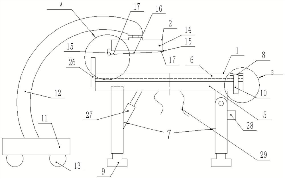
[0030] Such as Figure 1 ~ Figure 4 As shown, an interventional contrast imaging device includes a workbench 1, one end of the workbench 1 is provided with a contrast imaging device 2, the workbench 1 includes a support plate 5, and a work plate 6 is slidably arranged on the support plate 5, and the support plate The bottom surface of 5 is provided with the first support rod 7 around, the first support rod 7 of one end is plugged with the support plate 5, the first support rod 7 of the other end is hinged with the support plate 5, and the first support rod 7 of one end is plugged. A second hydraulic cylinder 27 is arranged on the top, and the telescopic end of the second hydraulic cylinder 27 is fixed to the support plate 5, and the lower end of the first support rod 7 is fixed to the first hydraulic cylinder 9. The contrast imaging device 2 includes a curved rod 12. One end of the curved rod 12 is connected to the base 11 with the first self-locking wheel 13, and the other en...

Embodiment 2

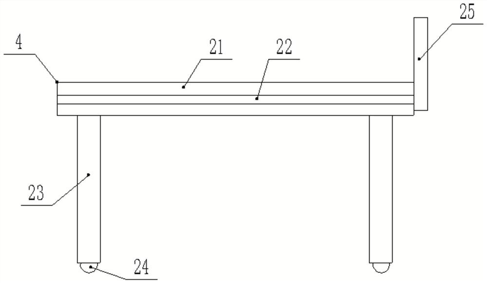
[0035] Preferably, also include delivery bed 4, delivery bed 4 comprises fixed plate 21, is provided with second support rod 23 around the bottom surface of fixed plate 21, is provided with universal wheel 24 under the second support bar 23, the both sides of fixed plate 21 A chute 22 is provided, and the fixed plate 21 and the supporting plate 5 share the same working plate 6 , and the chute 22 is used for sliding of the working plate 6 . The other end of the delivery bed 4 away from the slide-in end of the working plate 6 is fixed with a support bar 25 .

[0036] Setting the transport bed 4 matched with the working board 6 can further reduce the time required for transporting and moving the patient, so as to gain time for treatment.

[0037] Other structures of this embodiment are the same as those of Embodiment 1.

Embodiment 3

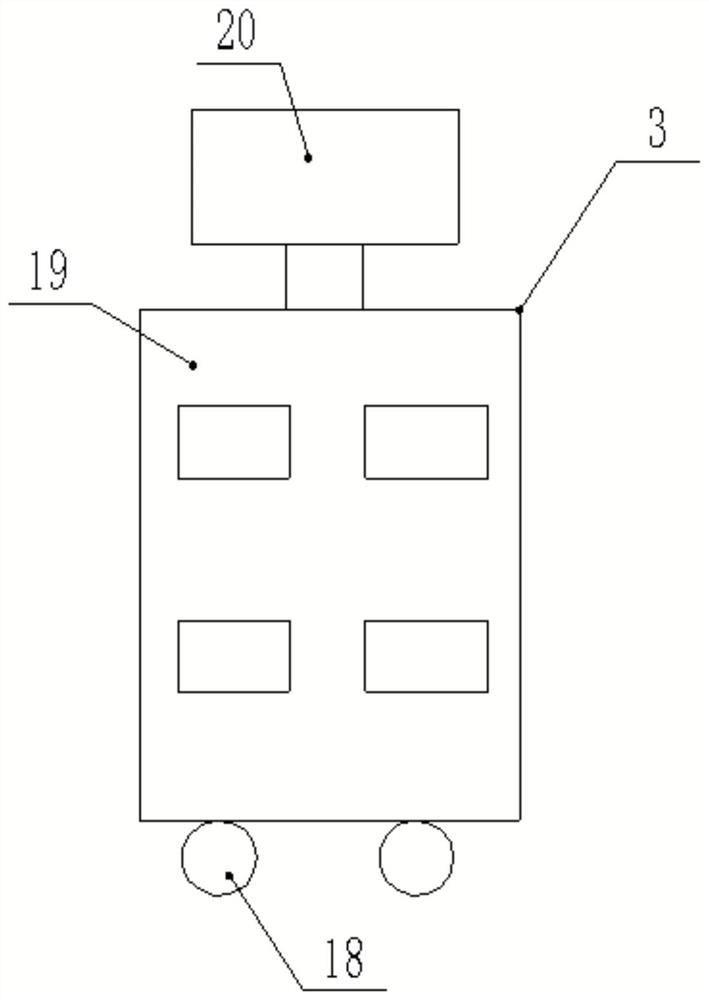
[0039] Preferably, it also includes a main controller 3, the main controller 3 includes a control panel 19, a display 20 is arranged on the control panel 19, a second self-locking wheel 18 is arranged under the control panel 19, and the contrast imaging main body 14 and the control panel 19 electrically connected.

[0040] Other structures of this embodiment are the same as those of Embodiment 1.


PUM


Abstract

The invention provides an interventional contrast imaging device and method, which are used for solving the problems that a patient is inconvenient to transport and the patient needs to change a bed body back and forth for examinations of different positions during a current contrast examination. The interventional contrast imaging device comprises a workbench; one end of the workbench is provided with a contrast imaging device; the workbench comprises a supporting plate; a working plate is arranged on the supporting plate in a sliding manner; first supporting rods are arranged on the periphery of the bottom face of the supporting plate; the lower end of each first supporting rod is fixed to a hydraulic cylinder; the contrast imaging device comprises a bent rod; one end of the bent rod is connected with a base with a first self-locking wheel, and the other end of the bent rod is rotationally provided with a contrast imaging main body; the contrast imaging main body extends to a position above the working plate; and a working face of the contrast imaging main body is provided with detachable covering cloth. The interventional contrast imaging device is reasonable in design and convenient to install; the sliding working plate is arranged, and a patient can be placed on the working plate and slide to the workbench, so that the problem that the patient is inconvenient to carry is solved, and precious time is won for treatment.

Description

technical field [0001] The invention relates to the technical field of medical devices, in particular to an interventional contrast imaging device and method. Background technique [0002] Angiography equipment has a good effect on the examination and treatment of cardiovascular and cerebrovascular diseases, and has gradually become an essential clinical diagnosis and treatment equipment. [0003] At present, when angiography is performed, the patient needs to be examined on a special examination bed, and the patient needs to be moved from other beds to the examination bed first, but because some patients are of high weight or serious condition, it is troublesome to carry, wasting time and delaying The treatment time is shortened; during interventional surgery, blood stains and other dirt may be splashed on the imaging subject, which will lead to imaging failure and inaccurate results, which will affect the diagnosis. If the imaging device is moved aside first, It will affe...

Claims


Application Information

Patent Timeline
Patent Type & Authority Applications(China)
IPC IPC(8): A61G13/00A61G13/10A61G1/02A61G1/04A61B6/00A61B6/04
CPCA61G13/00A61G13/10A61G1/02A61G1/04A61B6/00A61B6/0407A61B6/4405A61B6/44
Inventor 任衍乔郑传胜陈磊阚雪锋
Owner XIEHE HOSPITAL ATTACHED TO TONGJI MEDICAL COLLEGE HUAZHONG SCI & TECH UNIV
Why Eureka
Try Eureka