
Digital pathological image classification method and system based on superpixel segmentation and image convolution

A technology of superpixel segmentation and digital pathology, applied in image analysis, medical image, image enhancement, etc., can solve the problems of loss, difficulty in expressing histopathological relationship and histological features, ignoring the spatial relationship of microscopic cells, etc., to improve accuracy. rate effect

Pending Publication Date: 2021-08-27
FUDAN UNIV SHANGHAI CANCER CENT
View PDF0 Cites 4 Cited by
  • Summary
  • Abstract
  • Description
  • Claims
  • Application Information

AI Technical Summary

Problems solved by technology

However, the traditional CNN processes structured two-dimensional array images with pixel values. This structured two-dimensional array data is difficult to express the relationship and histological characteristics between cells and glands in histopathology. Neglecting the spatial relationship between microscopic cells will lose some key feature information to improve model performance

Method used


Image

  • Digital pathological image classification method and system based on superpixel segmentation and image convolution
  • Digital pathological image classification method and system based on superpixel segmentation and image convolution
  • Digital pathological image classification method and system based on superpixel segmentation and image convolution

Examples


Embodiment 1

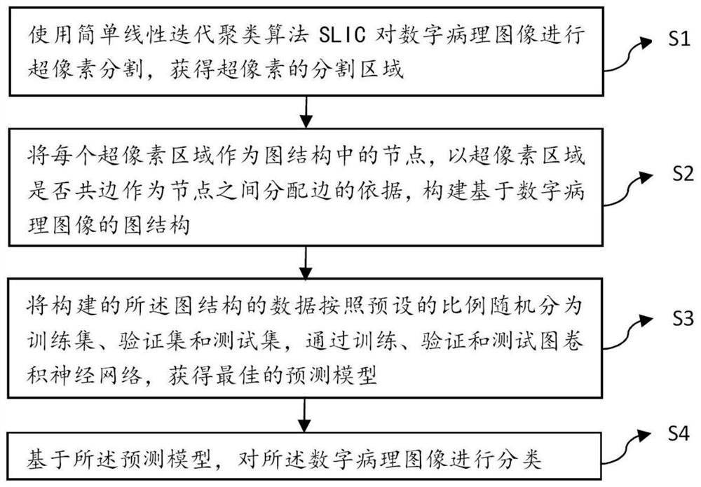
[0059] figure 1 It is a flowchart of a digital pathology image classification method based on superpixel segmentation and graph convolution. like figure 1 As shown, the present invention provides a kind of digital pathological image classification method based on superpixel segmentation and graph convolution, and described method comprises the following steps:

[0060] S1: Use the simple linear iterative clustering algorithm SLIC to perform superpixel segmentation on the digital pathology image, and obtain the superpixel segmentation area.

[0061] Specifically, step S1 includes:

[0062] S11: Initialize the seed point, that is, initialize the cluster center, and set the number of superpixels to be segmented;

[0063] S12: Reselect the seed point according to the gradient value of the pixel point in the n×n neighborhood of the seed point, so as to avoid the seed point falling on the edge with a larger gradient;

[0064] S13: assign a class label to each pixel point in the ...

Embodiment 2

[0096] Figure 7 It is a schematic diagram of the structure of a digital pathological image classification system based on superpixel segmentation and graph convolution. like Figure 7 As shown, the present invention also provides a digital pathological image classification system based on superpixel segmentation and graph convolution, said system comprising:

[0097] Segmentation module, for using simple linear iterative clustering algorithm SLIC to carry out superpixel segmentation to digital pathology image, obtain the segmentation area of ​​superpixel;

[0098] The construction module is used to use each superpixel area as a node in the graph structure, and whether the superpixel area is shared as the basis for allocating edges between nodes to construct a graph structure based on digital pathology images;

[0099] The training module is used to randomly divide the data of the graph structure constructed into a training set, a verification set and a test set according to...


PUM


Abstract

The invention relates to a digital pathological image classification method and system based on superpixel segmentation and image convolution, and the method comprises the following steps: S1, carrying out superpixel segmentation of digital pathological images through a simple linear iterative clustering algorithm SLIC, and obtaining a superpixel segmentation region; S2, taking each super-pixel region as a node in a graph structure, taking whether the super-pixel regions share the same edge as the basis of edge distribution between the nodes, and constructing the graph structure based on the digital pathological images; S3, randomly dividing the constructed data of the graph structure into a training set, a verification set and a test set according to a preset proportion, and obtaining an optimal prediction model by training, verifying and testing a graph convolutional neural network; and S4, based on the prediction model, classifying the digital pathological images. According to the invention, classification prediction of the digital pathological images is realized by using the graph convolutional neural network, and the accuracy of pathological image classification is improved.

Description

technical field [0001] The invention relates to the technical field of computer-aided diagnosis, in particular to a method and system for classifying digital pathological images based on superpixel segmentation and graph convolution. Background technique [0002] A standardized and unified pathology report provides sufficient information for clinicians, but the morphological evaluation of pathological indicators increases the workload of pathologists. Due to the maturity and standardization of pathological biopsy techniques, the number of pathological biopsy samples for diagnosis continues to rise, and the diagnostic results will be affected by factors such as fatigue and subjective experience. In recent years, the rapid development of digital pathological scanning technology and artificial intelligence (AI) has provided great potential for pathological diagnosis. Specifically, a deep convolutional neural network (CNN) is used to extract high-dimensional features in patholo...

Claims


Application Information

Patent Timeline
Patent Type & Authority Applications(China)
IPC IPC(8): G06K9/62G06N3/04G06N3/08G06T7/00G06T7/11G16H30/20
CPCG06T7/11G06N3/08G06T7/0012G16H30/20G06T2207/20081G06N3/045G06F18/24G06F18/214
Inventor 王奕张敬谊丁偕张伯强崔浩阳黄宗浩李渊张晖朱敏俊厉励张逸鲁高宇戴梅黄麒玮蔡云飞曹斌石强王正源王骏杰于镆铘崔敏杰
Owner FUDAN UNIV SHANGHAI CANCER CENT
Learn More