A single-bed PET delayed imaging method without concomitant CT radiation

An imaging method and CT image technology, applied in the fields of radiological diagnosis instruments, medical science, diagnosis, etc., can solve the problems of increasing the risk of cancer and increasing the radiation dose of patients, and achieve the goal of eliminating CT radiation and reducing stress. Effect

Active Publication Date: 2021-12-17
ZHEJIANG LAB +1
View PDF6 Cites 0 Cited by
  • Summary
  • Abstract
  • Description
  • Claims
  • Application Information

AI Technical Summary

Problems solved by technology

[0004] The original data required for PET imaging in PET / CT delayed scans come from the γ-rays produced by isotopic drugs. Adding delayed scans does not require the injection of additional isotopic drugs. However, since the delayed scan reconstruction algorithm includes attenuation correction, it is still necessary to acquire a patient’s CT This will increase the radiation dose received by patients, increase the risk of cancer for patients, and face greater medical ethics issues, which are important factors restricting PET delayed scan imaging

Method used


Image

  • A single-bed PET delayed imaging method without concomitant CT radiation
  • A single-bed PET delayed imaging method without concomitant CT radiation
  • A single-bed PET delayed imaging method without concomitant CT radiation

Examples


Embodiment 1

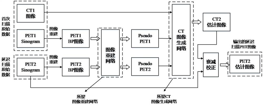
[0028] Such as figure 1 It is a schematic diagram of a single-bed PET delayed imaging method based on no accompanying CT radiation, and the method includes the following steps:

[0029] Step 1: Acquire a large amount of clinically normal and delayed PET / CT image data, including PET BP (Backprojection) and PET AC (attenuation correction) images of normal and delayed scans, as well as CT images of normal and delayed scans .

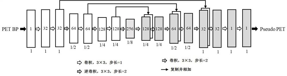
[0030] Step 2: Design and train PET image reconstruction supervised learning network;

[0031] In this embodiment, the image reconstruction network structure is as figure 2 As shown in , the input is a PET BP image (including normal scan and delayed scan image data), and the output is the predicted corresponding Pseudo PET image. The Pseudo PET image uses the PETAC image as a label, so that the image reconstruction network can convert the PET BP image into into PseudoPET images close to PET AC images. The objective function for training is expressed a...

Embodiment 2

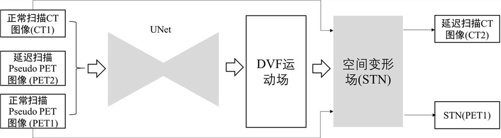
[0046] As a preferred solution, this embodiment considers that there are often clinically delayed scans with only one or more beds of PET data, and proposes a single-bed PET delayed imaging method without accompanying CT radiation. This method utilizes delayed scans The PET image is zero-filled, and the mask image is generated and the layer information image is set for registration to realize the CT image generation for attenuation correction. Figure 5 Shown is a schematic diagram of the mask image and the layer information image. The gray area in the normal scan image and the delayed scan image is the coordinate interval of the image along the Z axis. The scan area of ​​the normal scan is usually wider than the delay scan area, and the corresponding coordinate interval It is also bigger; the mask image is an image composed of 0 and 1, which sets the elements of the Z-axis coordinate interval of the delayed scanning image to 1, and sets the other areas to 0, and the mask image...


PUM


Abstract

The invention discloses a single-bed PET delayed imaging method without accompanying CT radiation. First, an image reconstruction network capable of converting a PET BP image into a pseudo PET image closer to a real PET image is used to obtain a normal scan and a delayed scan. The PET BP image is converted into a Pseudo PET image, and then a CT image generation network is used to input the normal scan Pseudo PET image and CT image, as well as the delayed scan Pseudo PET image, and output the deformation field between the normal scan and the delayed scan and The CT image at the time of the delayed scan, which is finally used for attenuation correction in the reconstruction of the delayed scan PET image, obtains a PET image with accurate SUV quantification and is used for tumor detection. The method of the invention can eliminate the CT radiation received by the patient in delayed scanning, reduce the physiological and psychological pressure of the patient, and promote the application of delayed PET imaging.

Description

technical field [0001] The invention relates to reconstruction attenuation correction of PET delayed scanning images, in particular to a single-bed PET delayed imaging method without accompanying CT radiation. Background technique [0002] PET (Positron Emission Tomography) delayed scan is an important basis for the early diagnosis of lung cancer, and it is of great significance to improve the cure rate of lung cancer and improve the quality of life of patients. According to the statistics of researchers, if early diagnosis and treatment of lung cancer patients can be carried out, the 5-year survival rate of lung cancer patients can be increased from 14% to 49%. In clinical practice, about 85% of lung cancer cases are advanced when diagnosed, and the 5-year survival rate of advanced lung cancer is less than 5%. Therefore, early detection, early diagnosis and early treatment are the key to improving the cure rate of lung cancer. PET imaging is a medical imaging that can prov...

Claims


Application Information

Patent Timeline
Patent Type & Authority Patents(China)
IPC IPC(8): A61B6/00A61B6/03
CPCA61B6/5235A61B6/037A61B6/032
Inventor 朱闻韬饶璠杨宝韩璐叶宏伟王瑶法
Owner ZHEJIANG LAB
Why Eureka
Try Eureka