Supercharge Your Innovation With Domain-Expert AI Agents!

Blood flow imaging method and ultrasonic imaging equipment

A blood flow and ultrasonic technology, applied in blood flow measurement devices, ultrasonic/sonic/infrasonic diagnosis, sonic diagnosis, etc., can solve the problems of missing key information, inaccuracy, low efficiency, etc.

Pending Publication Date: 2021-11-19
SHENZHEN MINDRAY BIO MEDICAL ELECTRONICS CO LTD
View PDF0 Cites 0 Cited by
  • Summary
  • Abstract
  • Description
  • Claims
  • Application Information

AI Technical Summary

Problems solved by technology

[0005] However, there are some differences between traditional color Doppler ultrasound imaging and ultrasound vector flow imaging in terms of operation and display. Senior medical personnel are already familiar with the operation of traditional color Doppler ultrasound. Unfamiliar; In addition, the diagnosis of many diseases is based on traditional color Doppler ultrasound. Due to the short development time of ultrasound vector flow imaging, the application of ultrasound vector flow imaging is still in its infancy. Medical personnel may use ultrasound vector flow imaging due to their experience Insufficient results in low efficiency or failure to find key information
Therefore, clinically, medical personnel mostly use traditional color Doppler ultrasound for auxiliary diagnosis, but the obtained blood flow information is relatively little or inaccurate

Method used


Image

  • Blood flow imaging method and ultrasonic imaging equipment
  • Blood flow imaging method and ultrasonic imaging equipment
  • Blood flow imaging method and ultrasonic imaging equipment

Examples


Embodiment Construction

[0049] Exemplary embodiments of the present invention will be described in more detail below with reference to the accompanying drawings. Although exemplary embodiments of the present invention are shown in the drawings, it should be understood that the invention may be embodied in various forms and should not be limited to the embodiments set forth herein. Rather, these embodiments are provided for more thorough understanding of the present invention and to fully convey the scope of the present invention to those skilled in the art.

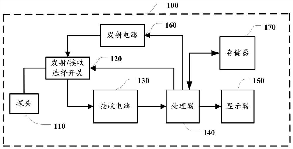
[0050] like figure 1Shown is a structural block diagram of an ultrasonic imaging device. Wherein, the ultrasonic imaging device 100 includes an ultrasonic probe 110 , a sending / receiving selection switch 120 , a receiving circuit 130 , a processor 140 , a display 150 , a transmitting circuit 160 and a memory 170 . The sending / receiving selection switch 120 can activate the ultrasonic probe 110 to transmit ultrasonic beams to the target area vi...


PUM


Abstract

The invention discloses a blood flow imaging method and ultrasonic imaging equipment. The method comprises the steps: emitting ultrasonic waves to a target blood flow area along a first scanning angle and a second scanning angle; receiving an ultrasonic echo returned from the target blood flow area to obtain a first ultrasonic echo signal and a second ultrasonic echo signal; performing signal processing on the first ultrasonic echo signal to obtain a first color ultrasound image and a first blood flow velocity; performing signal processing on the second ultrasonic echo signal to obtain a second blood flow velocity; synthesizing the first blood flow velocity and the second blood flow velocity to obtain a vector velocity of blood flow; and displaying the vector velocity of the blood flow and the first color ultrasound image in an overlapping manner. The vector velocity of the blood flow is superposed on the color ultrasound image to be displayed, so that the color ultrasound image printing habit of medical personnel is not changed, the traditional clinical diagnosis is not influenced, more blood flow information provided based on vector blood flow imaging is obtained, and more help is provided for auxiliary diagnosis and treatment of the medical personnel.

Description

technical field [0001] Embodiments of the present invention relate to the technical field of ultrasound imaging and display, and in particular to a blood flow imaging method and ultrasound imaging equipment. Background technique [0002] Color Doppler ultrasound, referred to as color Doppler ultrasound, generally uses autocorrelation technology for Doppler signal processing. The blood flow signal obtained by autocorrelation technology is color-coded and superimposed on the two-dimensional image in real time to form a color Doppler image. Ultrasound blood flow image. Color Doppler ultrasound examination is non-invasive, it can provide rich visual information of blood flow, and is widely used in clinical examination and qualitative analysis in practice. [0003] Traditional color Doppler ultrasound calculates the velocity component of the blood flow along the ultrasonic propagation direction, and the red and blue color spectrum in the color Doppler ultrasound only indicates t...

Claims


Application Information

Patent Timeline
IPC IPC(8): A61B8/06A61B8/00
CPCA61B8/06A61B8/4444A61B8/461A61B8/467A61B8/469A61B8/488
Inventor 杜宜纲范伟向兰茜王渊王凯张晟
Owner SHENZHEN MINDRAY BIO MEDICAL ELECTRONICS CO LTD
Features
  • R&D
  • Intellectual Property
  • Life Sciences
  • Materials
  • Tech Scout
  • Unparalleled Data Quality
  • Higher Quality Content
  • 60% Fewer Hallucinations
Learn More