
System and method of capturing and managing information during a medical diagnostic imaging procedure

Inactive Publication Date: 2005-03-24
MILLER LANDON C G
View PDF42 Cites 260 Cited by
  • Summary
  • Abstract
  • Description
  • Claims
  • Application Information

AI Technical Summary

Benefits of technology

[0014] One advantage of the present invention is that a system and method of capturing and managing data obtaining during a medical diagnostic imaging procedure is provided that improves the accuracy of diagnostic interpretation of the data. Another advantage of the present invention is that a system and method is provided that links information technology with medical diagnostic technology. Still another advantage of the present invention is that a system and method is provided that reduces the amount of time it takes to evaluate the data, since the data is provided in a consistent, organized manner. A further advantage of the present invention is that a system and method is provided that efficiently manages the diagnostic data so that associated costs are reduced. Still a further advantage of the present invention is that a system and method is provided that improves diagnostic accuracy and intervention through enhanced information. Still yet a further advantage of the present invention is that a system and method is provided for capturing the real-time impressions and observations of the test administrator.
[0015] Other features and advantages of the present invention will be readily understood as the same become better understood after reading the subsequent description when considered in connection with the accompanying drawings.

Problems solved by technology

Despite the extensive diagnostic capabilities of these imaging devices, however, there is no formal system and method available for managing the collection of the data and measurements derived during a test procedure, as well as the technician's impression.
While these imaging devices work well, a primary disadvantage is that the physician reviewing the data generated during a test procedure is merely reviewing discrete images or information obtained during a dynamic testing procedure in order to make a diagnosis.
Unfortunately, in the case of imaging procedures such as sonogram or ultrasound imaging, the discrete data may not be sufficient.
Additionally, there may be a significant amount of detailed data and measurements associated with an imaging procedure, such as a sonogram study.
In the past, these forms were cumbersome, expensive, and created opportunities for transcription error.
This situation also creates an opportunity for error, and generally slows down the overall diagnostic process and intervention.
Another disadvantage is that the forms may by incomplete, or restrictive due to space limitations.

Method used


Image

  • System and method of capturing and managing information during a medical diagnostic imaging procedure
  • System and method of capturing and managing information during a medical diagnostic imaging procedure
  • System and method of capturing and managing information during a medical diagnostic imaging procedure

Examples


Embodiment Construction

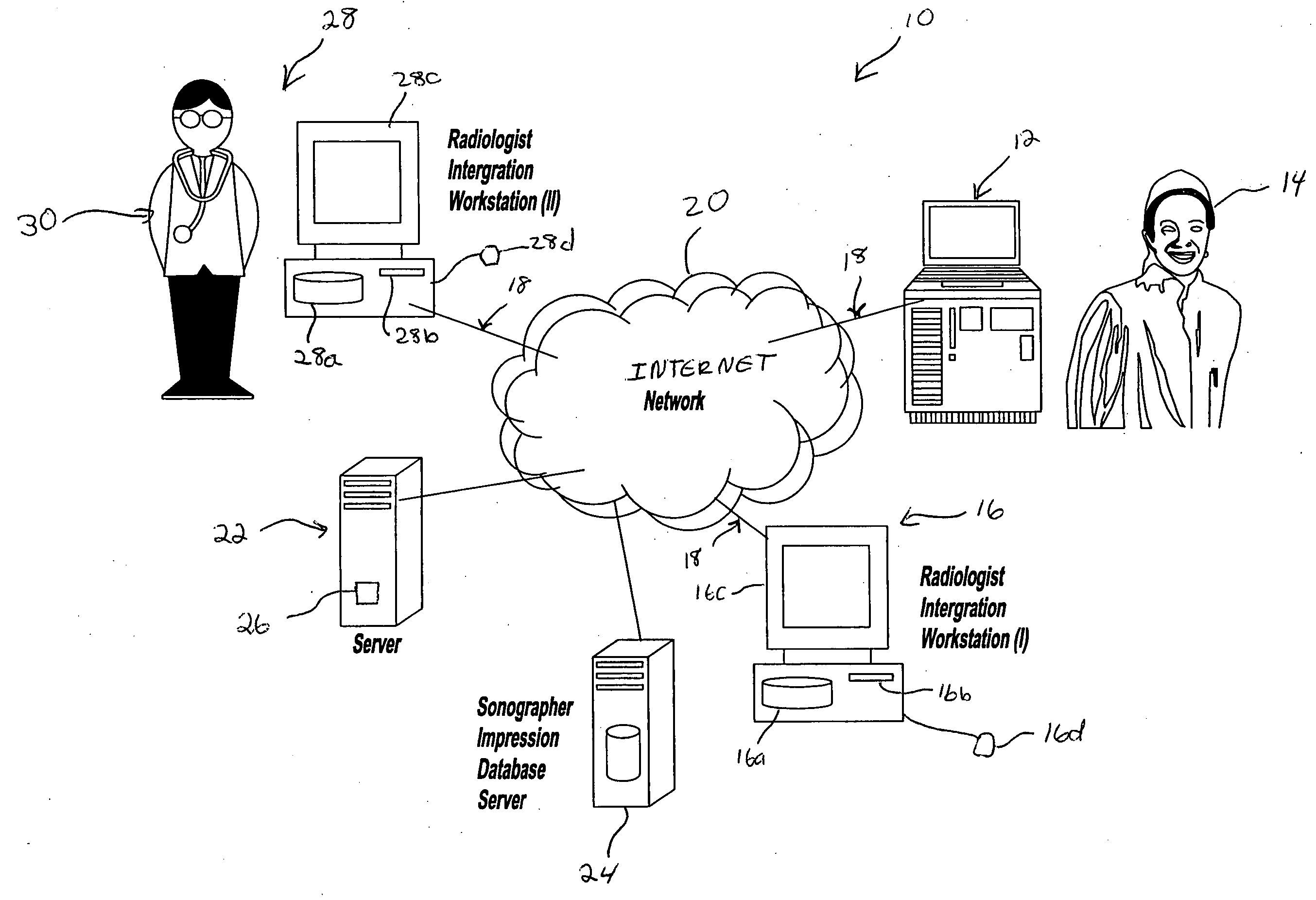
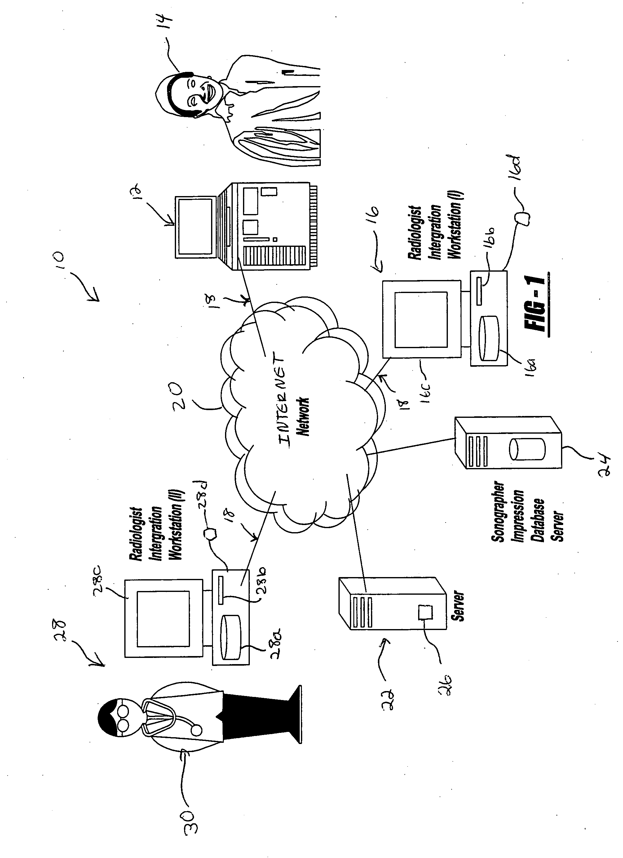
) Referring to FIG. 1, a system 10 is disclosed for capturing and managing data obtained during a medical diagnostic imaging procedure. It should be appreciated that in this example the imaging procedure is a sonogram performed using a sonographic device by a technician, referred to in the art as a sonographer. The system and method captures the impressions or observations of the ultrasound medical sonographer and the data upon which such impression is based, in real time, and stores the information on a computer system having a data storage means containing a database.

[0020] The system 10 includes an imaging device 12 operated by a technician 14. In this example, the imaging device 12 is an ultrasound imaging device that includes an ultrasonic transducer, a processor, a transmitter and a receiver. The ultrasonic transducer generates an ultrasonic signal that is transmitted to the processor. The processor collects the data and constructs a real-time image from the collected data. It...


PUM


Abstract

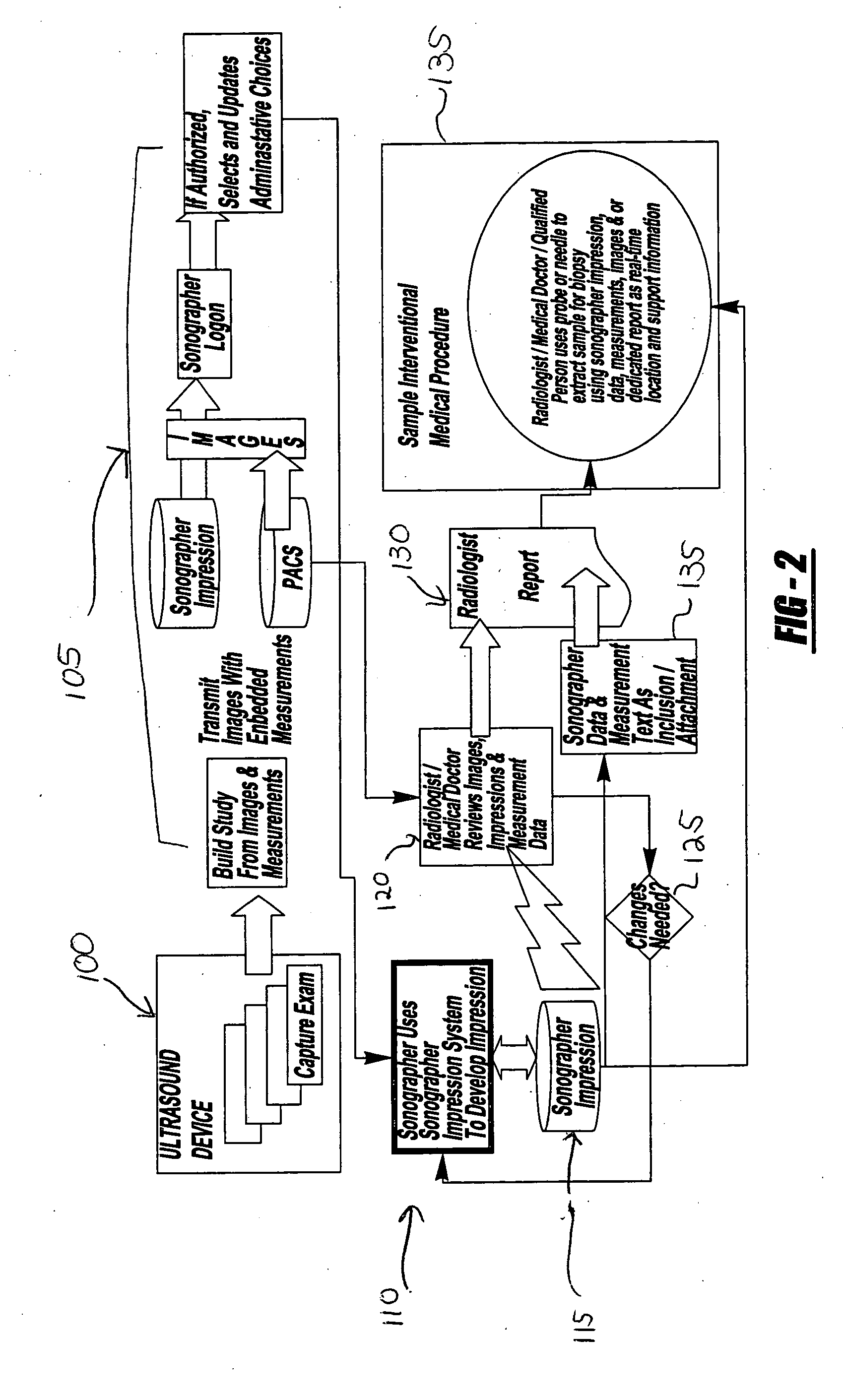
A system and method of capturing and managing the medical information obtained during an imaging procedure is provided. The system includes a sonogram imaging device operated by a technician, and a technician computer system in communication with the imaging device via a communications network. A computer server communicates with the technician computer system, and includes a medical diagnostic imaging software program. A healthcare provider computer system is operatively in communication with the computer server via the communications network. The method includes the steps of conducting the imaging procedure and building a study from the generated image and captured impression of the real-time observations of the generated image and measurement data by the technician. The study is stored in the database associated with a computer server and is accessible by a healthcare professional using a healthcare provider computer system for preparing a medical diagnosis.

Description

RELATED APPLICATION [0001] This application claims priority of U.S. Provisional Patent Application Ser. No. 60 / 501,620 filed Sep. 8, 2003, which is incorporated herein by reference.BACKGROUND OF THE INVENTION [0002] I. Field of the Invention [0003] The present invention relates generally to medical procedures and more particularly to a system and method of capturing and managing health information obtained during a medical diagnostic imaging procedure. [0004] II. Description of the Prior Art [0005] Advances in biomedical engineering have resulted in a proliferation of medical devices used for diagnostic purposes. In particular, various medical diagnostic imaging devices are well known in the art, such as magnetic resonance imaging (MRI), ultrasound, endoscopy, or computed tomography (CT). These imaging devices are usually standalone systems and are usually operated by highly trained technicians. [0006] Medical diagnostic imaging devices typically include a processor for processing t...

Claims


Application Information

Patent Timeline
IPC IPC(8): A61B5/00
CPCA61B5/0002
Inventor MILLER, LANDON C.G.
Owner MILLER LANDON C G
Learn More