Blood flow imaging method for 3D endoscope based on multi-angle scattering random matrix

A technology of random scattering and imaging method, applied in the fields of endoscopy, application, medical science, etc., can solve the problem of inability to achieve statistical separation of single and multiple scattered light intensity, and achieve the effect of accurate blood flow and good imaging effect.

Active Publication Date: 2021-12-03
SHANGHAI JIAO TONG UNIV
View PDF5 Cites 0 Cited by
  • Summary
  • Abstract
  • Description
  • Claims
  • Application Information

AI Technical Summary

Problems solved by technology

The signal recorded by the reflective laser speckle system contains both single-scattered and multiple-scattered photons, corresponding to the blood flow information of the surface and deeper blood vessels of the object during the operation. Statistical separation of light intensities

Method used


Image

  • Blood flow imaging method for 3D endoscope based on multi-angle scattering random matrix
  • Blood flow imaging method for 3D endoscope based on multi-angle scattering random matrix
  • Blood flow imaging method for 3D endoscope based on multi-angle scattering random matrix

Examples


Embodiment

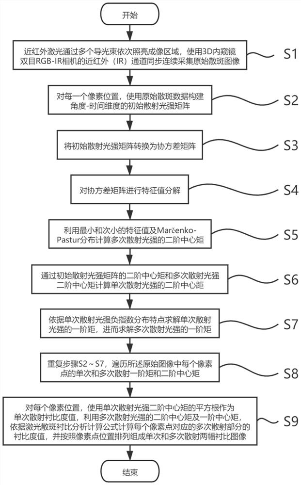
[0053] Such as figure 1 As shown, the present embodiment provides a 3D endoscopic blood flow imaging method based on a multi-angle scattering random matrix, which includes the following steps:

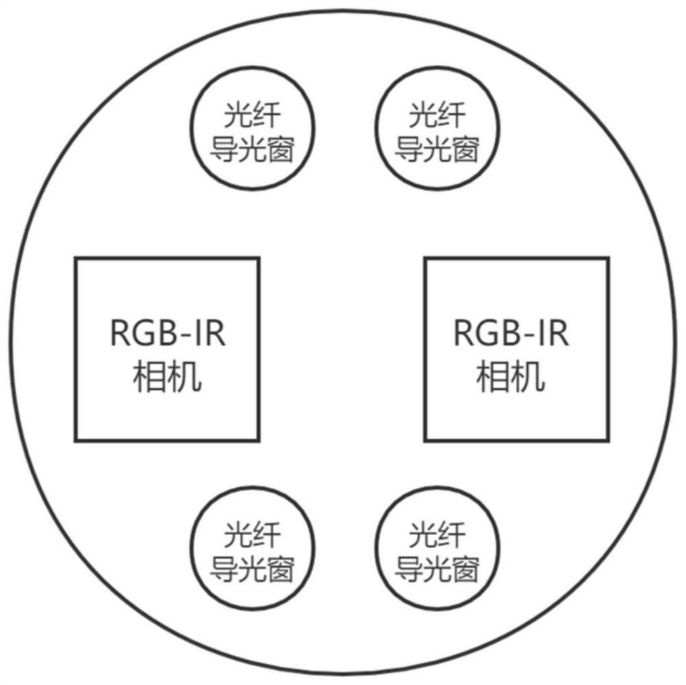
[0054] S1. Connect the near-infrared coherent laser light source to the n guide beams of the 3D endoscope, and control each guide beam to pass through in turn to form an illumination cycle. Use the near-infrared channels of the two cameras of the 3D endoscope to simultaneously collect the original scattered light. For speckle images, N=2n original speckle images are collected in one illumination cycle. Specifically: the near-infrared coherent laser light source is connected to the n beam guides of the 3D endoscope, and the light outlets of the n beam guides are distributed at fixed positions at the front end of the 3D endoscope. With different angle relationships, the light guides take turns to emit light in one lighting cycle. The near-infrared channels of the two cameras are used to...


PUM


Abstract

The invention relates to a blood flow imaging method for a 3D endoscope based on a multi-angle scattering random matrix, and the method comprises the following steps: S1, connecting a light source to n light guide beams of the 3D endoscope, controlling each light guide beam to pass light in sequence to form an illumination period, synchronously collecting original speckle images by using near-infrared channels of two cameras of the 3D endoscope, and collecting 2n original speckle images in one illumination period; S2, constructing an initial scattering light intensity matrix for any pixel position; S3, acquiring a single-scattering light intensity second-order central moment, a multiple-scattering light intensity second-order central moment and a multiple-scattering light intensity first-order central moment of a pixel point; S4, traversing all the pixel points; and S5, acquiring a single-scattering contrast degree value and a multiple-scattering contrast degree value of each pixel point, and arranging according to the positions of the pixel points to form a single-scattering contrast degree image and a multiple-scattering contrast degree image. Compared with the prior art, more accurate surface and deep blood flow information can be obtained.

Description

technical field [0001] The invention relates to the technical field of optical imaging, in particular to a blood flow imaging method for 3D endoscope based on multi-angle scattering random matrix. Background technique [0002] 3D endoscopy is an emerging tool in surgery that can provide real-time depth perception. Through three-dimensional reconstruction of the contour data of internal tissues, organs or lesions in the human body, 3D endoscopy effectively improves the accuracy, feasibility and scientificity of the current diagnosis and treatment process. At present, 3D endoscopes have been introduced and used in various departments such as abdominal cavity, cranial cavity, and gastroenterology. [0003] At present, intravenous injection of indocyanine green dye combined with fluorescence imaging has been used to obtain visual images of blood flow in tumors and vasculature, but this technique is subject to the metabolic cycle of dye injection and is prone to false positive r...

Claims


Application Information

Patent Timeline
Patent Type & Authority Applications(China)
IPC IPC(8): A61B1/00A61B1/04A61B1/07A61B5/00
CPCA61B1/04A61B1/07A61B1/00193A61B1/00009A61B1/00064A61B1/00131A61B5/0086
Inventor 苗鹏
Owner SHANGHAI JIAO TONG UNIV
Why Eureka
Try Eureka