Brachial plexus ultrasound image segmentation method, system, medium and device based on U-Net

An ultrasound image and brachial plexus technology, applied in the field of brachial plexus ultrasound image segmentation, can solve problems such as adding steps, and achieve the effects of noise suppression, high speed and good effect

Pending Publication Date: 2021-12-03
JINLING INST OF TECH
View PDF3 Cites 0 Cited by
  • Summary
  • Abstract
  • Description
  • Claims
  • Application Information

AI Technical Summary

Problems solved by technology

Most methods preprocess ultrasound images to remove noise, which adds unnecessary steps to the model training process

Method used


Image

  • Brachial plexus ultrasound image segmentation method, system, medium and device based on U-Net
  • Brachial plexus ultrasound image segmentation method, system, medium and device based on U-Net

Examples


Embodiment Construction

[0032] In order to enable those skilled in the art to better understand the solution of the present application, the technical solution in the embodiment of the application will be clearly and completely described below in conjunction with the accompanying drawings in the embodiment of the application. Obviously, the described embodiment is only It is an embodiment of a part of the application, but not all of the embodiments. Based on the embodiments in this application, all other embodiments obtained by persons of ordinary skill in the art without creative efforts shall fall within the scope of protection of this application.

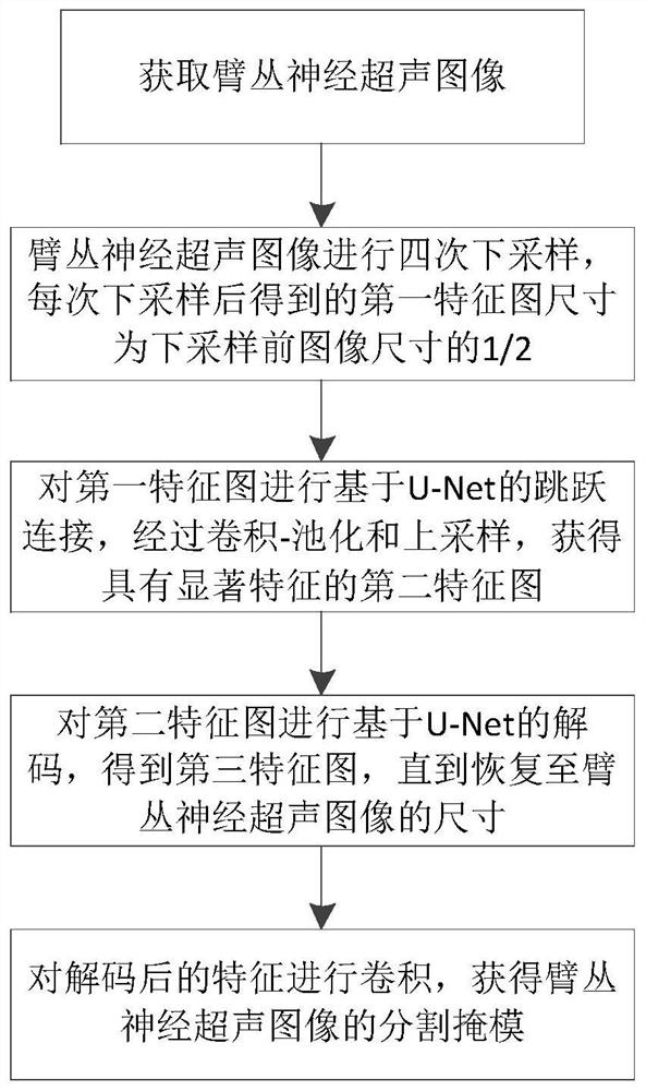
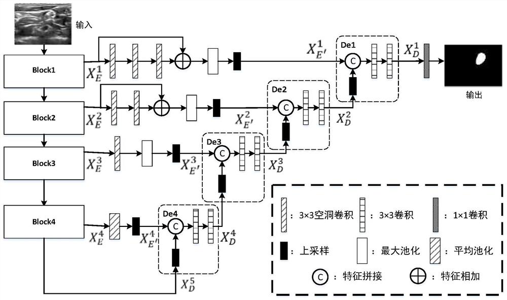
[0033] Convolutional neural network based on deep learning is currently a popular image segmentation technology. Most methods preprocess ultrasound images to remove noise, which adds unnecessary steps to the model training process, because the deep network can adaptively learn the main features. . Although the U-Net-based model takes into account the ...


PUM


Abstract

The invention discloses a segmentation method, system, medium and device based on a U-Net network. The method comprises the following steps: acquiring a brachial plexus ultrasound image; performing down-sampling on the brachial plexus ultrasound image for four times to obtain a first feature map of which the size is 1/2 of the size of the image before down-sampling; performing U-Net-based jump connection on the first feature map to obtain a second feature map with significant features; decoding the second feature map based on U-Net, and recovering to the size of the brachial plexus ultrasound image; and carrying out convolution on the decoded features to obtain a segmentation mask of the brachial plexus ultrasound image. According to the invention, no operator is used for image preprocessing, and the image segmentation process is simplified; the features are extracted by using an efficient OfficientNet series network, so that the speed is high and the effect is good; U-Net jump connection is designed, more significant features are extracted on a large receptive field by using cavity convolution and maximum pooling operation, and the effect of noise suppression is achieved.

Description

technical field [0001] The invention belongs to the technical field of computer vision and artificial intelligence, in particular to a U-Net-based brachial plexus ultrasound image segmentation method, system, medium and device. Background technique [0002] Medical image processing has become an important part of digital medicine. With the help of image segmentation, the useful part of the image can be found for doctors. Ultrasound scanning is a widely used form of medical imaging that detects various diseases and injuries by diagnosing and analyzing structures such as muscles and nerves inside the human body. Therefore, how to automatically segment ultrasound images to obtain regions of interest is of great significance. [0003] Compared with high-contrast CT and MRI, automatic segmentation of ultrasound images is a more challenging task. During the formation of brachial plexus (Brachial Plexus, BP) ultrasound images, due to random disturbances of electronic devices, the...

Claims


Application Information

Patent Timeline
Patent Type & Authority Applications(China)
IPC IPC(8): G06T7/00G06N3/04G06N3/08G06T5/00G06T7/10
CPCG06T7/0012G06T7/10G06T5/002G06N3/08G06T2207/10132G06T2207/20021G06T2207/20081G06T2207/20084G06T2207/30004G06N3/045
Inventor 孔令军王茜雯包云超侯文涛刘伟光周耀威闫佳艺
Owner JINLING INST OF TECH
Why Eureka
Try Eureka