Ultrasonic contrast imaging method and ultrasonic imaging device for fallopian tube and storage medium

A technique of fallopian tube and angiography, applied in the field of ultrasound imaging, can solve the problems of missing fallopian tubes, difficult to locate the spatial position, etc., and achieve the effect of improving the efficiency of diagnosis

Pending Publication Date: 2021-12-21
SHENZHEN MINDRAY BIO MEDICAL ELECTRONICS CO LTD +1
View PDF0 Cites 0 Cited by
  • Summary
  • Abstract
  • Description
  • Claims
  • Application Information

AI Technical Summary

Problems solved by technology

At present, most of the ultrasonic four-dimensional salpingography imaging can only display four-dimensional contrast or tissue images alone. When users make a diagnosis, they often need to combine the image information and the relative spatial relationship of the two, especially for some abnormal conditions (missing fallopian tubes, blockage, unobstructed), it is difficult to locate the spatial position only by relying on contrast image information

Method used


Image

  • Ultrasonic contrast imaging method and ultrasonic imaging device for fallopian tube and storage medium
  • Ultrasonic contrast imaging method and ultrasonic imaging device for fallopian tube and storage medium
  • Ultrasonic contrast imaging method and ultrasonic imaging device for fallopian tube and storage medium

Examples


Embodiment Construction

[0020] In order to make the objects, technical solutions and advantages of the present invention more apparent, exemplary embodiments according to the present invention will be described in detail below with reference to the accompanying drawings. Apparently, the described embodiments are only some embodiments of the present invention, rather than all embodiments of the present invention, and it should be understood that the present invention is not limited by the exemplary embodiments described here. Based on the embodiments of the present invention described in the present invention, all other embodiments obtained by those skilled in the art without creative effort shall fall within the protection scope of the present invention.

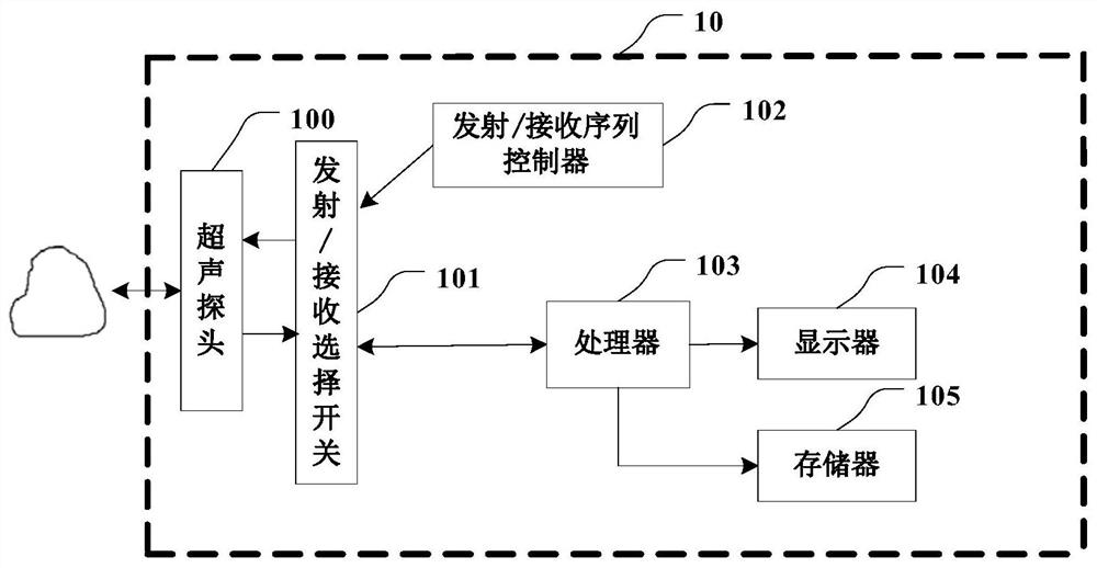
[0021] First, refer to figure 1 An exemplary ultrasonic measurement device for implementing the ultrasound-enhanced ultrasound imaging method of the fallopian tube according to the embodiment of the present application will be described.

[0022] ...


PUM


Abstract

The invention provides a ultrasonic contrast imaging method and ultrasonic imaging device for a fallopian tube and a storage medium. The method comprises the following steps: acquiring four-dimensional contrast data and four-dimensional tissue data of a fallopian tube area of a target object; respectively acquiring three-dimensional radiography data and three-dimensional tissue data at at least one moment from the four-dimensional radiography data and the four-dimensional tissue data; rendering the acquired three-dimensional radiography data and the three-dimensional tissue data at each moment, and fusing rendering results obtained after rendering the three-dimensional radiography data and the three-dimensional tissue data at the same moment to obtain a mixed rendered image; performing feature extraction on the mixed rendering image, and outputting an analysis result of the fallopian tube region based on a feature extraction result; and displaying the mixed rendered image and the analysis result. According to the oviduct ultrasound contrast imaging method and device, a user can understand and observe the spatial position relation and the flowing condition of the contrast agent in the tissue more visually.

Description

technical field [0001] The present application relates to the technical field of ultrasonic imaging, and more specifically relates to a fallopian tube ultrasonic contrast imaging method, an ultrasonic imaging device and a storage medium. Background technique [0002] Ultrasound instruments are generally used by doctors to observe the internal tissue structure of the human body. The doctor places the operating probe on the skin surface corresponding to the part of the human body to obtain an ultrasonic image of the part. Due to its safety, convenience, non-destructive, and low-cost characteristics, ultrasound has become the main auxiliary means of diagnosis for doctors. [0003] Ultrasound contrast agent is a substance used to enhance the image contrast in ultrasound imaging. It is generally a micron-scale diameter coated microbubble. The microbubble has a strong acoustic impedance and enters the blood circulation system through intravenous injection to enhance the reflection...

Claims


Application Information

Patent Timeline
Patent Type & Authority Applications(China)
IPC IPC(8): G06T7/00G06T5/50G06T15/08G06T15/50G06T15/06
CPCG06T7/0014G06T5/50G06T15/08G06T15/506G06T15/06G06T2207/10132G06T2207/20221G06T2210/41
Inventor 张新玲王艾俊林穆清邹耀贤何绪金
Owner SHENZHEN MINDRAY BIO MEDICAL ELECTRONICS CO LTD
Why Eureka
Try Eureka