Blood vessel segmentation method, terminal and computer readable storage medium

A technology of blood vessels and microvessels, which is applied in the field of medical assistance, can solve the problems affecting the judgment of doctors, uneven illumination, and blurred images, and achieve the effects of insensitive image brightness and image contrast, avoiding image quality problems, and good segmentation effects

Inactive Publication Date: 2021-12-21
WUHAN ENDOANGEL MEDICAL TECH CO LTD
View PDF9 Cites 5 Cited by
  • Summary
  • Abstract
  • Description
  • Claims
  • Application Information

AI Technical Summary

Problems solved by technology

[0003] Due to the constraints of hardware conditions and the level of endoscopists, there are phenomena such as uneven illumination, blurred images, and weak contrast between microvessels and image background in the magnified imaging of gastric mucosa staining, which makes it impossible to perform well on the magnified images of gastric mucosa staining. Vascular segmentation, thus affecting the judgment of follow-up physicians and causing misdiagnosis

Method used


Image

  • Blood vessel segmentation method, terminal and computer readable storage medium
  • Blood vessel segmentation method, terminal and computer readable storage medium
  • Blood vessel segmentation method, terminal and computer readable storage medium

Examples


Embodiment Construction

[0129]The following will clearly and completely describe the technical solutions in the embodiments of the application with reference to the drawings in the embodiments of the application. Apparently, the described embodiments are only some of the embodiments of the application, not all of them. Based on the embodiments in this application, all other embodiments obtained by those skilled in the art without making creative efforts belong to the scope of protection of this application.

[0130] In the description of the present application, it should be understood that the terms "center", "longitudinal", "transverse", "length", "width", "thickness", "upper", "lower", "front", " The orientation or positional relationship indicated by "rear", "left", "right", "vertical", "horizontal", "top", "bottom", "inner", "outer", etc. is based on the orientation shown in the drawings Or positional relationship is only for the convenience of describing the present application and simplifying ...


PUM


Abstract

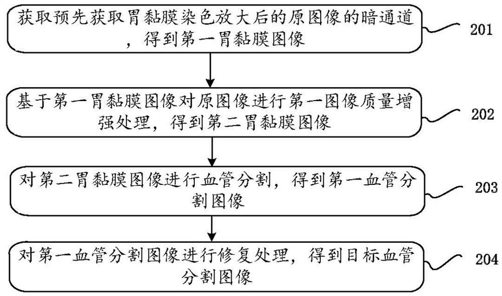
The invention provides a blood vessel segmentation method, a terminal and a computer readable storage medium. The method comprises the steps: obtaining a dark channel of an original image obtained after gastric mucosa is dyed and amplified in advance, and obtaining a first gastric mucosa image; performing first image quality enhancement processing on the original image based on the first gastric mucosa image to obtain a second gastric mucosa image; performing blood vessel segmentation on the second gastric mucosa image to obtain a first blood vessel segmentation image; and repairing the first blood vessel segmentation image to obtain a target blood vessel segmentation image. According to the method, the requirement for the quality of a gastroscope image is not high, the method is not sensitive to the image brightness and the image contrast, a good segmentation effect is achieved, further, the segmented image is repaired, the accuracy of the image is improved, and the situation that a doctor misdiagnoses due to the image quality problem is avoided.

Description

technical field [0001] The present application relates to the field of medical assistance technology, and in particular to a blood vessel segmentation method, a terminal, and a computer-readable storage medium. Background technique [0002] Microvessel analysis of gastric mucosal staining and magnification imaging plays an important role in the diagnosis of early gastric cancer: Yao Kenfumi pointed out in the VS typing theory that there are irregular microvessels with demarcation lines in early gastric cancer; Yagi Kazuyoshi in mesh-loop It is pointed out in the theory that the microvessels of differentiated gastric cancer can be divided into network structure and ring structure under the state of staining amplification, and the vascular morphology of undifferentiated gastric cancer can be divided into Lemon boat shape, wave shape and spiral shape. The analysis of microvessels in the above theory is a qualitative analysis based on personal experience. If quantitative analysi...

Claims


Application Information

Patent Timeline
Patent Type & Authority Applications(China)
IPC IPC(8): G06T7/10G06T7/68G06T5/00G06T5/20G06T5/30
CPCG06T7/10G06T7/68G06T5/002G06T5/005G06T5/20G06T5/007G06T5/30G06T5/003G06T2207/20028G06T2207/20032G06T2207/20192G06T2207/30101G06T2207/10068
Inventor 李昊胡珊于红刚
Owner WUHAN ENDOANGEL MEDICAL TECH CO LTD
Why Eureka
Try Eureka