Retinal vascular image segmentation method and system based on differential attention

A retinal blood vessel and image segmentation technology, used in image analysis, image enhancement, image data processing, etc., can solve the problems of complex network, inability to extract small blood vessel parts, and unsatisfactory segmentation effect, achieve accurate boundary area and solve segmentation problems. The effect of insufficient precision and accurate segmentation results

Pending Publication Date: 2022-01-04
SHANDONG NORMAL UNIV
View PDF0 Cites 0 Cited by
  • Summary
  • Abstract
  • Description
  • Claims
  • Application Information

AI Technical Summary

Problems solved by technology

Although these methods perform well, they inevitably make the network more complex. When the feature accumulation reaches the limit, it may not be possible to extract small blood vessel parts, resulting in unsatisfactory segmentation results.

Method used


Image

  • Retinal vascular image segmentation method and system based on differential attention
  • Retinal vascular image segmentation method and system based on differential attention
  • Retinal vascular image segmentation method and system based on differential attention

Examples


Embodiment 1

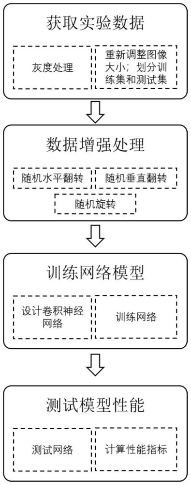
[0033] like figure 1 As shown, the present embodiment provides a method for segmenting retinal blood vessel images based on differential attention, which specifically includes the following steps:

[0034] S101: Acquire retinal blood vessel images;

[0035] S102: Based on the retinal blood vessel image and the multi-scale residual network based on differential attention, obtain the retinal fundus blood vessel image segmentation result;

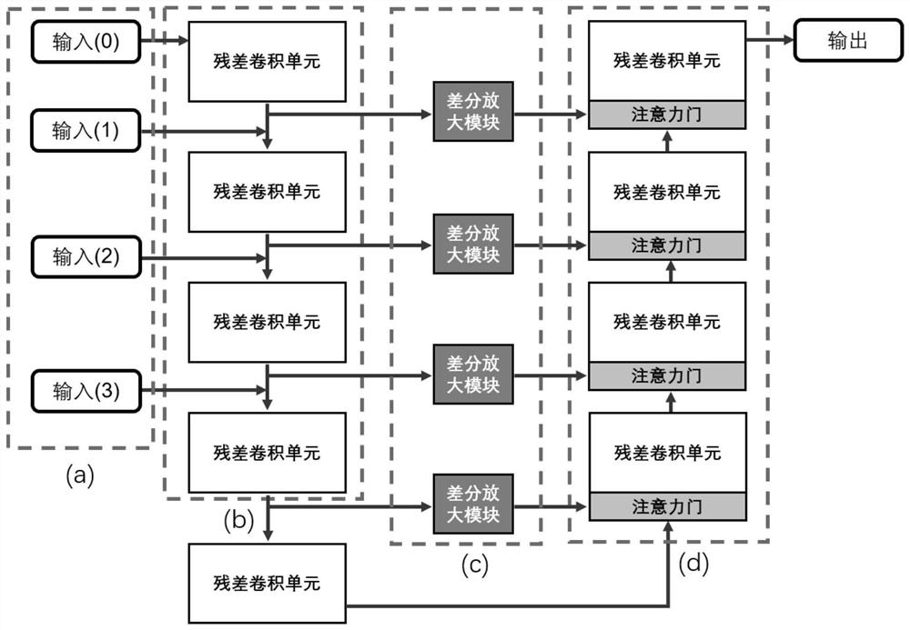
[0036] Among them, such as figure 2 As shown, the multi-scale residual network based on differential attention includes a multi-scale input module, an encoder module, a differential amplification module and a decoder module; the multi-scale input module is used to extract multi-scale information of retinal blood vessel images; the encoder module It is used to encode multi-scale information; the differential amplification module is used to extract the low-frequency information and high-frequency information of the encoded multi-scale informa...

Embodiment 2

[0068] This embodiment provides a retinal vessel image segmentation system based on differential attention, which specifically includes:

[0069] An image acquisition module, which is used to acquire retinal blood vessel images;

[0070] The image segmentation module is used to obtain the retinal fundus vascular image segmentation result based on the retinal vascular image and the multi-scale residual network based on differential attention;

[0071] Wherein, the multi-scale residual network based on differential attention includes a multi-scale input module, an encoder module, a differential amplification module and a decoder module; the multi-scale input module is used to extract multi-scale information of retinal blood vessel images; the encoder module uses It is used to encode multi-scale information; the differential amplification module is used to extract the low-frequency information and high-frequency information of the encoded multi-scale information respectively, and...

Embodiment 3

[0074] This embodiment provides a computer-readable storage medium, on which a computer program is stored. When the program is executed by a processor, the steps in the method for segmenting retinal blood vessel images based on differential attention as described above are implemented.


PUM


Abstract

The invention belongs to the field of medical image segmentation, and provides a retinal vascular image segmentation method and system based on differential attention. The method comprises the following steps: acquiring a retinal vascular image; and obtaining a retinal fundus vascular image segmentation result based on the retinal vascular image and a differential attention-based multi-scale residual network, wherein the differential attention-based multi-scale residual network comprises a multi-scale input module, an encoder module, a differential amplification module and a decoder module; the multi-scale input module is used for extracting multi-scale information of the retinal vascular image; the encoder module is used for encoding the multi-scale information; the differential amplification module is used for extracting low-frequency information and high-frequency information of the encoded multi-scale information and then extracting features of the low-frequency information and the high-frequency information; and an attention mechanism is introduced into the decoder module, so that an attention degree of an area needing to be paid attention to is improved, irrelevant areas are inhibited, and the extracted low-frequency and high-frequency features are finally restored to original resolutions.

Description

technical field [0001] The invention belongs to the field of medical image segmentation, and in particular relates to a retinal blood vessel image segmentation method and system based on differential attention. Background technique [0002] The statements in this section merely provide background information related to the present invention and do not necessarily constitute prior art. [0003] As a non-invasive rapid diagnosis method for modern ophthalmic diseases, retinal fundus vessel segmentation is an important part of computer-aided clinical diagnosis of retinal diseases. It can help ophthalmologists diagnose certain retinopathy, such as retinal vascular occlusion, diabetic retinopathy, high blood pressure and glaucoma. The characteristics of these diseases can be obtained by observing changes in retinal blood vessels, such as shape, width, branching pattern, etc. Early detection and diagnosis can help potential patients to carry out effective prevention and treatment...

Claims


Application Information

Patent Timeline
Patent Type & Authority Applications(China)
IPC IPC(8): G06T7/11G06N3/04G06N3/08
CPCG06T7/11G06N3/08G06T2207/10032G06T2207/20081G06T2207/20084G06T2207/20192G06T2207/30041G06T2207/30101G06N3/045
Inventor 李登旺张焱黄浦王婷赵睿孙晓蕾陈美荣吴冰
Owner SHANDONG NORMAL UNIV
Why Eureka
Try Eureka