Capsule endoscope
A technology of capsule endoscope and capsule shell, which is applied in the fields of endoscopy, medical science, surgery, etc., and can solve the problems of large axial length of the capsule shell, reduced image accuracy, and large intestinal error.
- Summary
- Abstract
- Description
- Claims
- Application Information
AI Technical Summary
Problems solved by technology
Method used
Image
Examples
Embodiment 1
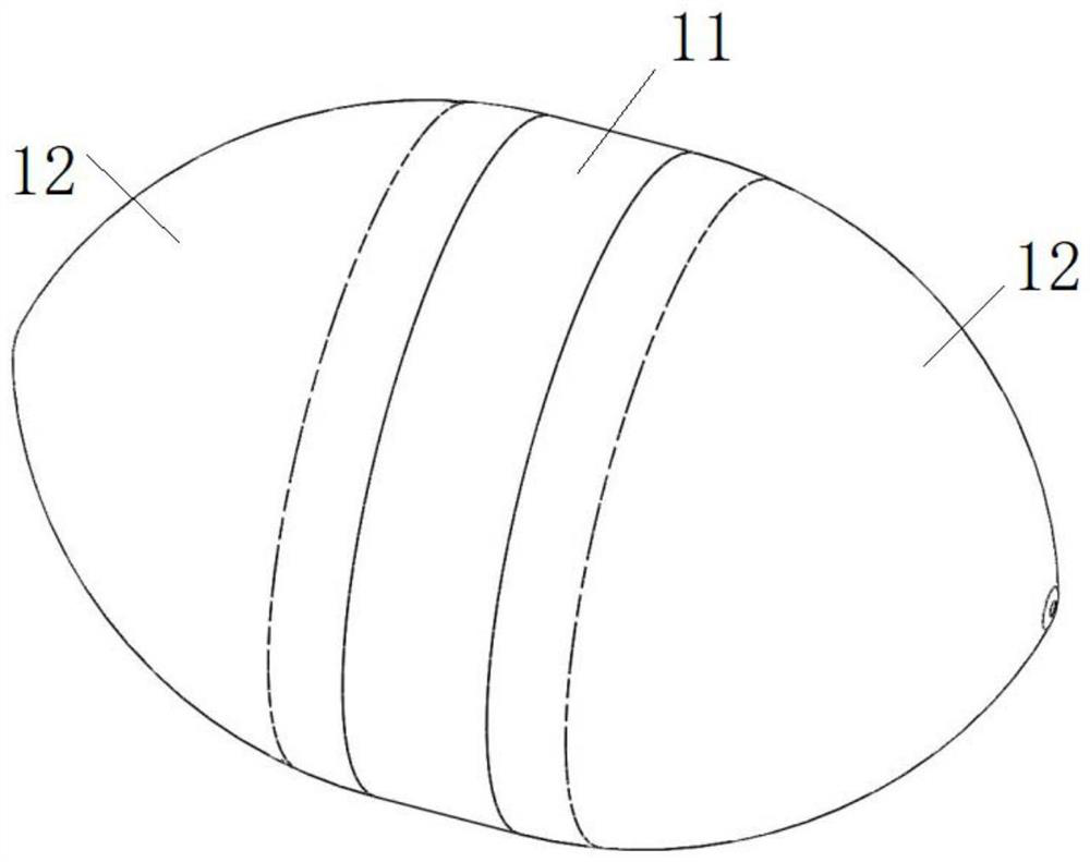
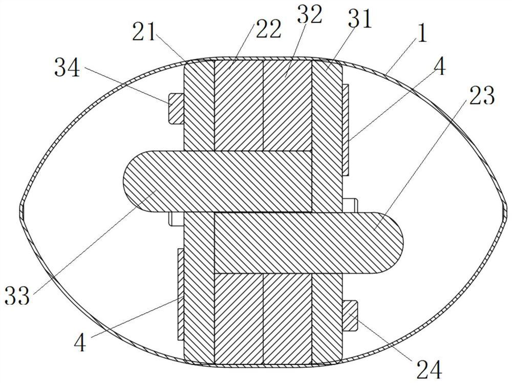
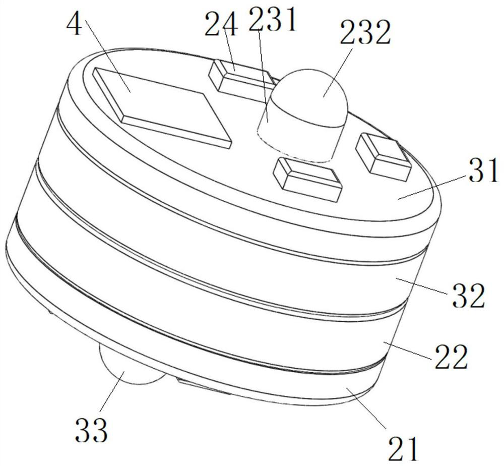
[0054] This embodiment provides a capsule endoscope, such as Figure 1 to Figure 6 As shown, it includes a capsule shell 1 and two sets of image acquisition mechanisms.
[0055] Such as figure 1 and figure 2 As shown, the capsule shell 1 has an installation cavity. Optionally, the capsule shell 1 includes a middle shell 11 and a transparent shell 12 detachably arranged on openings at both ends of the middle shell 11 . For example, the transparent shell 12 is sleeved outside the opening of the middle shell 11, and is connected by a buckle, or threaded, or directly socketed, or other existing detachable connection methods. The transparent shell 12 forms an observation window, which is also convenient for the lens to pass through the transparent shell 12 to collect images of the intestinal tract; of course, the middle shell 11 can also be made of a transparent material. Optionally, as in figure 1 As shown, the capsule shell 1 is in the shape of a rugby ball, or a capsule of ...
Embodiment 2
[0079] This embodiment provides a capsule endoscope, such as Figure 7 and Figure 8 As shown, it is compared with the capsule endoscope provided in embodiment 1, the difference is:
[0080] The structures of the power supply components are different. The first power supply component 22 and the second power supply component 32 are not provided with the above-mentioned relief holes and installation holes, and the first power supply component 22 and the second power supply component 32 are still arranged in the first space. At this time, the first power supply component 22 is clamped between the first circuit board 21 and the second circuit board 31, the second power supply component 32 is clamped between the first circuit board 21 and the second circuit board 31, and the second power supply component 32 is clamped between the first circuit board 21 and the second circuit board 31. A power supply part 22 and a second power supply part 32 are arranged away from the first body 23...
Embodiment 3
[0084] This embodiment provides a capsule endoscope. Compared with the capsule endoscopes provided in Embodiment 1 and Embodiment 2, the difference lies in:
[0085] The first lens 232 is located outside the first circuit board 21, the second lens 332 is located outside the second circuit board 31, and the projections of the first body 231 and the second body 331 on the above-mentioned cross section in the first space are along the capsule shell. There is an overlapping portion in the radial direction, the first body 231 no longer passes through the second circuit board 31, the second body 331 no longer passes through the first circuit board 21, the first power supply part 22 and the second power supply part 32 are set In the first space, the projections of the two power supply components on the above-mentioned cross-section may have no or overlapping parts along the radial direction of the capsule shell, and the axial length of the two sets of image acquisition mechanisms can ...
PUM
 Abstract
Description
Claims
Application Information
 - R&D
- Intellectual Property
- Life Sciences
- Materials
- Tech Scout
- Unparalleled Data Quality
- Higher Quality Content
- 60% Fewer Hallucinations
Browse by: Latest US Patents, China's latest patents, Technical Efficacy Thesaurus, Application Domain, Technology Topic, Popular Technical Reports.
© 2025 PatSnap. All rights reserved.Legal|Privacy policy|Modern Slavery Act Transparency Statement|Sitemap|About US| Contact US: help@patsnap.com


