
A suture-free coagulation-assisted fixation cardiac patch and preparation method thereof

A heart and patch technology, which is applied in medical science, ligaments, prostheses, etc., can solve the problems of large trauma, weak patch fit, easy slipping, etc., so as to reduce postoperative complications and improve myocardial repair effect , the effect of reducing the difficulty of surgery

Active Publication Date: 2022-07-29
ZHEJIANG UNIV
View PDF6 Cites 0 Cited by
  • Summary
  • Abstract
  • Description
  • Claims
  • Application Information

AI Technical Summary

Problems solved by technology

[0003] Most of the implantation methods of existing cardiac patches are suture and bonding, and the suture method has problems such as complicated operation process, large trauma, easy postoperative complications and long recovery time (recorded in literature 1 (An Elastic, Biodegradable Cardiac Patch Induces Contractile Smooth Muscle and Improves CardiacRemodeling and Function in Subacute Myocardial Infarction[J].Journal of the American College of Cardiology,2007,49(23):2292-2300)), there is a problem that the patch is not firmly attached and is easy to slip Problem (recorded in literature 2 (Engineering a naturally-derived adhesive and conductive cardiopatch[J].Biomaterials,2019,207) and literature 3(Flexible shape-memory scaffold for minimally invasive delivery of functional tissues[J].Nature Materials,2017) )
Adding a microneedle array on the side of the patch facing the heart has been proven to assist in the fixation of the patch. The shape of the microneedles is mostly conical without barbs, and the base of the patch is mostly smooth (recorded in literature 4 Cardiaccell–integrated microneedle patch for treating myocardial infarction[J].Science Advances,2018,4(11)), when the base of this heart patch is working, there is still a gap between it and the myocardium, which cannot be fully fitted

Method used


Image

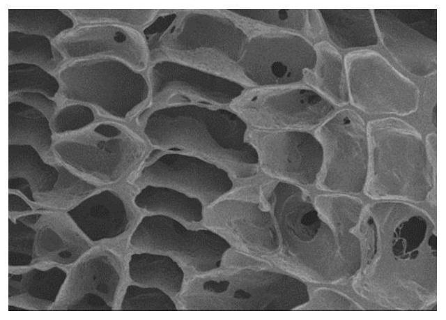
  • A suture-free coagulation-assisted fixation cardiac patch and preparation method thereof
  • A suture-free coagulation-assisted fixation cardiac patch and preparation method thereof
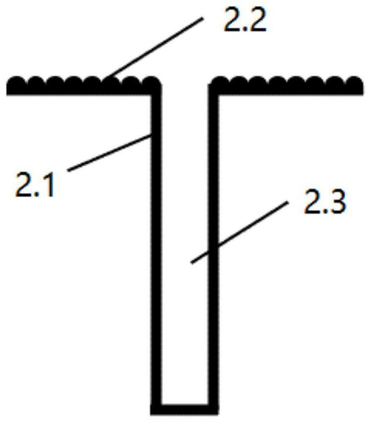
  • A suture-free coagulation-assisted fixation cardiac patch and preparation method thereof

Examples


Embodiment 1

[0078] A preparation method of a suture-free coagulation-assisted fixation heart patch, comprising the following steps:

[0079] Raw material preparation:

[0080] Barb microneedle: the diameter of the microneedle is 0.4 mm, and the angle between the bevel tip-shaped bevel and the axial direction of the microneedle is 30°; the cross-sectional shape of the barb is triangular; three groups of barbs are distributed along the longitudinal direction of the microneedle. Barbs, each group of barbs consists of 3 barbs, and the angle between the barbs and the microneedles is 30°; the barb microneedles are made of polylactic acid.

[0081] The force when the barbed microneedles were inserted into the chicken breast after being pulled out from the chicken breast was measured by a universal mechanical testing machine (Instron 5543A), and it was 0.122N.

[0082] (1) Model A is 3D printed, and the model A includes a base, and a plurality of cylinders and spherical recessed structures are f...

Embodiment 2

[0088] A preparation method of a suture-free coagulation-assisted fixation heart patch, comprising the following steps:

[0089] Raw material preparation:

[0090] Barb microneedle: the diameter of the microneedle is 0.5 mm, and the angle between the bevel tip-shaped bevel and the axial direction of the microneedle is 30°; the cross-sectional shape of the barb is triangular; 4 groups of barbs are distributed along the longitudinal direction of the microneedle. Barbs, each group of barbs consists of 3 barbs, and the angle between the barbs and the microneedles is 30°; the barb microneedles are made of polycaprolactone.

[0091] The force when the barbed microneedle was inserted into the chicken breast after being pulled out from the chicken breast was measured by a universal mechanical testing machine (Instron 5543A), and it was 0.200N.

[0092] Porogen: Use a sieve to screen out gelatin microspheres with a diameter of 100 μm.

[0093] (1) By 3D printing model C, the model C ...

Embodiment 3

[0099] A preparation method of a suture-free coagulation-assisted fixation heart patch, comprising the following steps:

[0100] Raw material preparation:

[0101] Barb microneedle: the diameter of the microneedle is 0.4 mm, and the angle between the bevel tip-shaped bevel and the axial direction of the microneedle is 30°; the cross-sectional shape of the barb is square; 5 groups of barbs are distributed along the longitudinal direction of the microneedle. Barbs, each group of barbs is composed of 3 barbs, and the angle between the barbs and the microneedles is 30°; the barb microneedles are made of polypropylene.

[0102] The force when the barbed microneedle was inserted into the chicken breast after being pulled out from the chicken breast was measured by a universal mechanical testing machine (Instron 5543A), and it was 0.220N.

[0103] (1) By 3D printing the model E, the model E includes a base, and several cylinders are formed on one side of the base, and the several cy...


PUM

PropertyMeasurementUnit
angleaaaaaaaaaa
lengthaaaaaaaaaa
pore sizeaaaaaaaaaa

Abstract

The invention relates to a suture-free coagulation-assisted fixation heart patch and a preparation method thereof. The cardiac patch comprises a polymer-promoting coagulation base, barbed microneedles fixed on the polymer-promoting coagulation base, and a polymer coagulation-promoting base tape. The side with the microneedles has a microporous structure that does not penetrate the solid polymer base; the barbed microneedles are microneedles with barbs, and the angle α between the microneedles and the plane where the polymer procoagulant base is located is 60-90 °, the microneedles are evenly distributed on the polymer procoagulant substrate, and the distribution density is 5-50 pieces / cm 2 ; the barbs are located on the curved surface of the microneedles and face the polymeric procoagulant substrate. The preparation method adopts the method of mold porosity, porogen porosity or thermal-induced phase separation porosity, and combines barbed microneedles to obtain a suture-free coagulation-assisted fixation heart patch while forming a microporous structure. The method is simple, and the prepared heart patch has excellent therapeutic effect.

Description

technical field [0001] The invention belongs to the technical field of biomedical materials, and relates to a suture-free coagulation-assisted fixation heart patch and a preparation method thereof. Background technique [0002] Myocardial infarction is a relatively common and fatal type of cardiovascular disease, and the main cause is myocardial necrosis caused by myocardial hypoxia-ischemia. Compared with traditional drugs, cell therapy and other methods, cardiac patch inhibits left ventricular remodeling, slows heart failure, and maintains cardiac function by providing mechanical support for infarcted myocardium. Combined with drug therapy. [0003] The existing implantation methods of cardiac patches are mostly suture and adhesive, and the suture methods have problems such as complicated surgical procedures, large trauma, easy postoperative complications, and long recovery time (recorded in document 1 (An Elastic, Biodegradable Cardiac Patch Induces). Contractile Smooth...

Claims


Application Information

Patent Timeline
Patent Type & Authority Patents(China)
IPC IPC(8): A61F2/08A61F2/00
CPCA61F2/0063A61F2/08A61F2/0811A61F2002/0847A61F2002/0817
Inventor 朱旸鲁育文高长有任探琛沈励胤洪晓倩
Owner ZHEJIANG UNIV
Learn More