
Eureka
For R&D, Eureka makes reading and utilizing patents & technical documents easy.
Eureka AIR
Designed for self-driven R&D workflows. Generate viable solutions, solve complex R&D challenges, empower your innovation with AI.

Eureka Materials
Designed for material experts only. Revolutionize your material R&D, from search, analyze, to developing new materials.

TechResearch
Generate reliable direction feasibility study reports for your R&D in just a few steps.

TechSeek
Discover and master advanced knowledge NOW. Basics, ideas, possibilities, all at once.

TechMind
As an expert in R&D Theories, TechMind can generates customized viable solutions instantly.

TechRisk
Analyze your overall solution with one click, know your potential R&D risks in advance.

TechMonitor
Get weekly tech updates, stay abreast of the latest tech innovations and key insights.
Intelligent judgment method for pulmonary fibrosis
A determination method and fibrosis technology, applied in the field of medical image analysis, can solve the problems of waste of training and testing time, difficulty of image block integration, and redundancy of target regions, so as to reduce training time, reduce time and storage overhead, and reduce calculation. amount of effect
- Summary
- Abstract
- Description
- Claims
- Application Information
AI Technical Summary
Problems solved by technology
Method used
Image

Examples
Embodiment 1
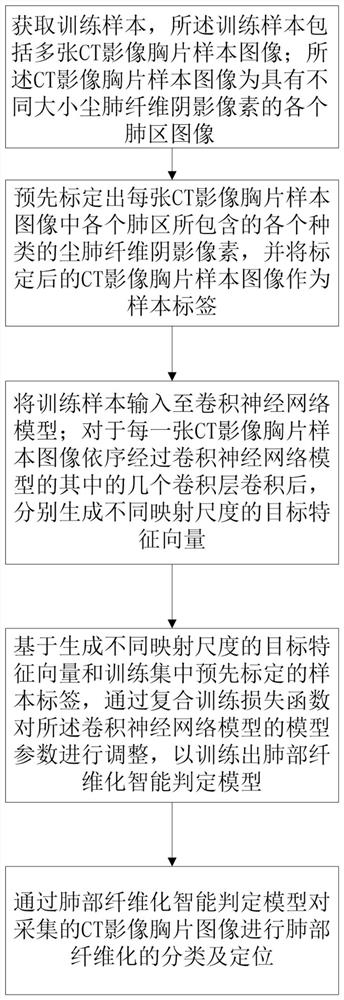
[0044] Please combine figure 1, a method for intelligently judging pulmonary fibrosis, comprising the following steps:
[0045] S1. Obtain a training sample, the training sample includes a plurality of CT image chest X-ray sample images; the CT image chest X-ray sample images are images of various lung regions with shadow pixels of pneumoconiosis fibers of different sizes;
[0046] S2. Pre-mark the shadow pixels of various types of pneumoconiosis fibers contained in each lung area in each CT image chest film sample image, and use the calibrated CT image chest film sample image as a sample label;
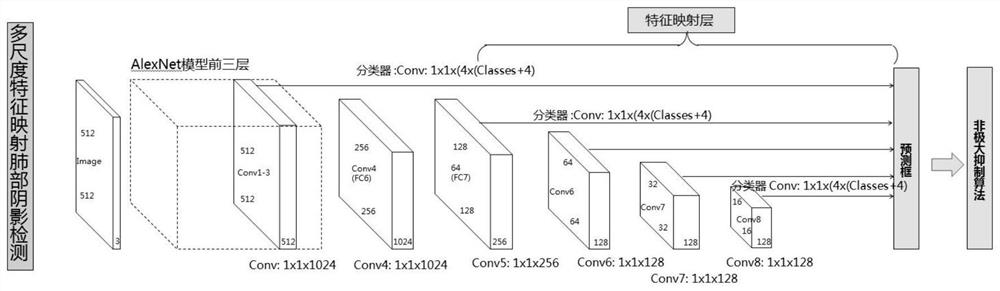
[0047] S3. Input the training samples into the convolutional neural network model; for each CT image chest X-ray sample image, after being convoluted by several convolutional layers of the convolutional neural network model, targets of different mapping scales are respectively generated Feature vector;
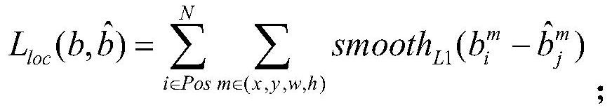
[0048] S4. Based on the generated target feature vectors of different mapping ...
Embodiment 2
[0069] This embodiment provides an intelligent determination device for pulmonary fibrosis using the method for intelligent determination of pulmonary fibrosis in Embodiment 1, including:
[0070] A sample acquisition module, which is used for training samples and includes a plurality of CT image chest film sample images; the CT image chest film sample images are images of various lung regions with different sizes of pneumoconiosis fiber shadow pixels;
[0071] A sample calibration module, which is used to pre-mark the various types of pneumoconiosis fiber shadow pixels contained in each lung area in each CT image chest film sample image, and use the calibrated CT image chest film sample image as a sample label;
[0072] A sample training module, which is used to input training samples to the convolutional neural network model; for each piece of CT image chest film sample image, after sequentially passing through several convolutional layer convolutions of the convolutional neu...
Embodiment 3
[0076] This embodiment provides a computer-readable storage medium, on which a computer program is stored. When the program is executed by a processor, the steps of the method for intelligent determination of pulmonary fibrosis in the above-mentioned embodiment 1 are realized.
PUM
 Abstract
Description
Claims
Application Information
 - R&D Engineer
- R&D Manager
- IP Professional
- Industry Leading Data Capabilities
- Powerful AI technology
- Patent DNA Extraction
Browse by: Latest US Patents, China's latest patents, Technical Efficacy Thesaurus, Application Domain, Technology Topic, Popular Technical Reports.
© 2024 PatSnap. All rights reserved.Legal|Privacy policy|Modern Slavery Act Transparency Statement|Sitemap|About US| Contact US: help@patsnap.com