Pre-positioning device for ultrasonic detection and implementation method thereof

A pre-positioning device, ultrasonic detection technology, applied in the direction of organ movement/change detection, ultrasonic/sonic wave/infrasonic wave diagnosis, ultrasonic/sonic wave/infrasonic wave Permian technology, etc. It is inconvenient to set up non-woven fabrics and other problems

Inactive Publication Date: 2022-04-05
PINGMEI SHENMA MEDICAL GRP GENERAL HOSPITAL
View PDF0 Cites 0 Cited by
  • Summary
  • Abstract
  • Description
  • Claims
  • Application Information

AI Technical Summary

Problems solved by technology

[0005] The purpose of the present invention is to provide a pre-positioning device for ultrasonic detection and its implementation method, which solves the problem that in the background art, during musculoskeletal detection, the patient's unconscious movements or twists will affect the detection effect, resulting in aggravated detection difficulty. Moreover, it is inconvenient

Method used


Image

  • Pre-positioning device for ultrasonic detection and implementation method thereof
  • Pre-positioning device for ultrasonic detection and implementation method thereof
  • Pre-positioning device for ultrasonic detection and implementation method thereof

Examples


Embodiment approach

[0046] In order to further and better explain the above-mentioned embodiments, the present invention also provides an embodiment, a method for implementing a pre-positioning device for ultrasonic testing, including the following steps:

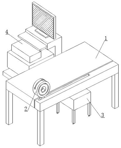
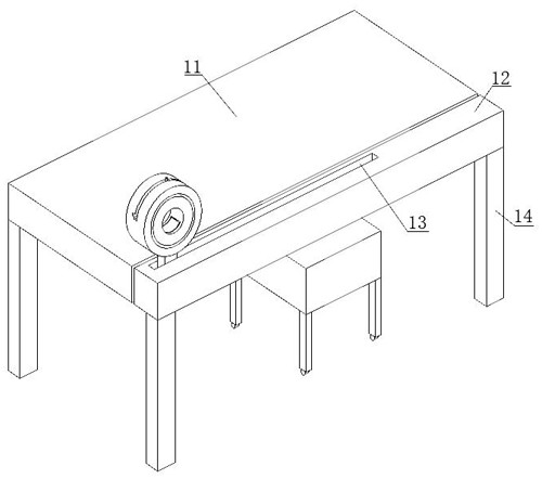
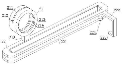
[0047] Step 1: When the patient needs to perform musculoskeletal ultrasound examination, push the component circle 21 to move from one end of the fixed bar 12 to the middle position of the fixed bar 12. At this time, the movement of the component circle 21 will make the transmission belt 22 rotate. Thereby, the seat 3 is pushed out through the linkage structure, so that its position is on the side of the hospital bed 1;

[0048] Step 2: The patient pulls one end of the disposable non-woven fabric 2292 from the bottom of the hospital bed 1, so that the disposable non-woven fabric 2292 covers the upper side of the seat 3, and the patient sits on the upper side of the seat 3, and ultrasonic testing will be required The part extends into the insid...


PUM


Abstract

The invention discloses a pre-positioning device for ultrasonic detection and an implementation method thereof, belongs to the technical field of muscle-bone ultrasonic detection, and aims to solve the problems that when an existing device is used for muscle-bone detection, limb parts of a patient are inconvenient to position and control, the detection effect can be influenced by unconscious actions or twisting of the patient, the detection difficulty degree is increased, and the detection time is shortened. The problem that non-woven fabrics are inconvenient to arrange one by one when a traditional device is used for carrying out muscle-bone ultrasonic detection is solved. According to the device, the component circular block is moved to a designated position, a detected part is clamped in the clamping groove, a doctor can conveniently detect the detected part by utilizing the rotatability of the rotary circular block in the component circular block, and when the component circular block moves, the seat covered with disposable non-woven fabric is pushed out through the linkage structure, so that a patient sits, and the detection efficiency is improved. The limb part of a patient is positioned and controlled, the detection effect is prevented from being affected by unconscious action or twisting of the patient, and health protection is achieved through disposable non-woven fabric.

Description

technical field [0001] The invention relates to the technical field of musculoskeletal ultrasonic testing, in particular to a pre-positioning device for ultrasonic testing and an implementation method thereof. Background technique [0002] Musculoskeletal ultrasonography can assist in the diagnosis of lesions in muscles, tendons, ligaments, bones and cartilage, joints, and nerves. It can also check for superficial soft tissue masses. In addition, it can also check for bone-related motor dysfunction pain and trauma. Tendon, ligament and peripheral nerve lesions, and musculoskeletal ultrasound detection is usually detected with a musculoskeletal ultrasound detector. [0003] When patients undergo musculoskeletal ultrasound testing, they need to show the detected parts to the medical staff. The medical staff use the musculoskeletal ultrasound detector to perform ultrasonic testing on the detected parts. Positioning control, the patient's unconscious movement or twisting will a...

Claims


Application Information

Patent Timeline
IPC IPC(8): A61B8/08A61B8/00A61G13/00A61G13/10A61G13/12A61G15/00A61G15/10A61G15/12
Inventor 孙婕
Owner PINGMEI SHENMA MEDICAL GRP GENERAL HOSPITAL
Why Eureka
Try Eureka