Medical image segmentation method and device, computer equipment and storage medium

A medical image and sample image technology, applied in the field of image processing, can solve problems such as blurred boundary information, difficult segmentation, and segmentation accuracy to be improved, and achieve the effect of accurate segmentation and improved segmentation performance

Active Publication Date: 2022-04-29
SHENZHEN UNIV
View PDF9 Cites 4 Cited by
  • Summary
  • Abstract
  • Description
  • Claims
  • Application Information

AI Technical Summary

Problems solved by technology

[0004] The purpose of the present invention is to provide a medical image segmentation method, device, computer equipment and storage medium, aiming to solve the problem of segmentation difficulty and segmentation accuracy rate that need to be improved due to blurred boundary information in the prior art

Method used


Image

  • Medical image segmentation method and device, computer equipment and storage medium
  • Medical image segmentation method and device, computer equipment and storage medium
  • Medical image segmentation method and device, computer equipment and storage medium

Examples


Embodiment Construction

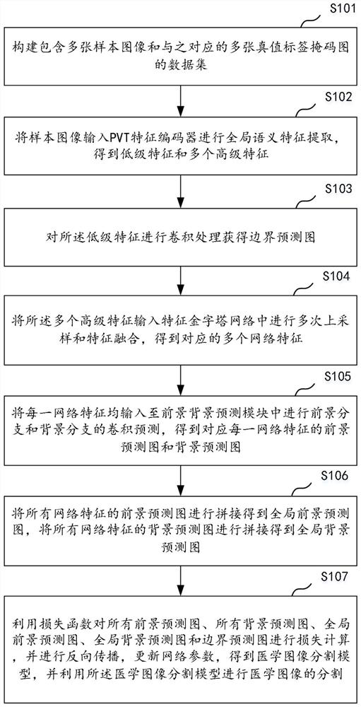
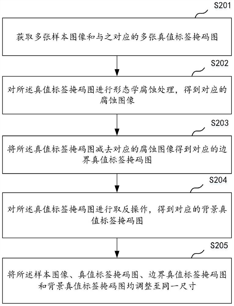
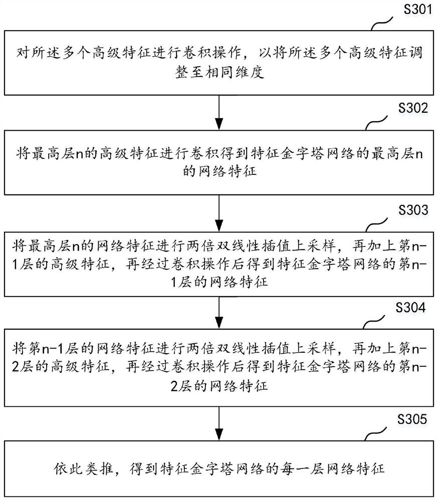
[0033] The following will clearly and completely describe the technical solutions in the embodiments of the present invention with reference to the accompanying drawings in the embodiments of the present invention. Obviously, the described embodiments are some of the embodiments of the present invention, but not all of them. Based on the embodiments of the present invention, all other embodiments obtained by persons of ordinary skill in the art without making creative efforts belong to the protection scope of the present invention.

[0034] It should be understood that when used in this specification and the appended claims, the terms "comprising" and "comprises" indicate the presence of described features, integers, steps, operations, elements and / or components, but do not exclude one or Presence or addition of multiple other features, integers, steps, operations, elements, components and / or collections thereof.

[0035] It should also be understood that the terminology used ...


PUM


Abstract

The invention discloses a medical image segmentation method and device, computer equipment and a storage medium. The method comprises the following steps: inputting a sample image into a PVT feature encoder for global semantic feature extraction to obtain a low-level feature and a plurality of high-level features; performing convolution processing on the low-level features to obtain a boundary prediction map; inputting the plurality of advanced features into a feature pyramid network for multiple times of up-sampling and feature fusion to obtain a plurality of corresponding network features; inputting each network feature into a foreground and background prediction module to obtain a foreground prediction map and a background prediction map; splicing the foreground prediction images to obtain a global foreground prediction image, and splicing the background prediction images to obtain a global background prediction image; and carrying out loss calculation by using the loss function, carrying out back propagation, and updating network parameters to obtain a medical image segmentation model. According to the invention, boundary information is used to guide feature expression, and a correction mechanism of foreground and background prediction difference is used to realize more accurate segmentation.

Description

technical field [0001] The invention relates to the technical field of image processing, in particular to a medical image segmentation method, device, computer equipment and storage medium. Background technique [0002] As one of the most common tasks in the field of medical image processing, medical image segmentation can accurately locate lesions and organ structures in the image and separate them from the image, providing important information for clinicians to perform further diagnosis or surgical treatment. clue. However, medical image segmentation is still a challenging task due to the characteristics of medical images such as high noise, low resolution and contrast, and complex and diverse imaging principles. [0003] In the prior art, medical image segmentation methods usually use manually designed features to identify the segmentation target in the image. This type of method needs to design different features for different lesion characteristics, and is easily inte...

Claims


Application Information

Patent Timeline
IPC IPC(8): G06T7/00G06V10/26G06V10/80G06V10/82G06K9/62G06N3/04G06N3/08
CPCG06T7/0012G06N3/084G06N3/045G06F18/253
Inventor 岳广辉卓桂彬李思莹周天薇汪天富段绿茵
Owner SHENZHEN UNIV
Why Eureka
Try Eureka