

Three-dimensional MRI focus image segmentation method and system
An image segmentation and lesion technology, applied in the field of lesion identification, can solve the problems of doctors’ professional requirements, easy missed detection, increase of doctor’s workload, etc., and achieve the effect of improving expression accuracy and auxiliary accuracy
- Summary
- Abstract
- Description
- Claims
- Application Information
AI Technical Summary
Problems solved by technology
Method used
Image

Examples
Embodiment Construction
[0066] The following will clearly and completely describe the technical solutions in the embodiments of the present invention with reference to the accompanying drawings in the embodiments of the present invention. Obviously, the described embodiments are only some, not all, embodiments of the present invention. Based on the embodiments of the present invention, all other embodiments obtained by persons of ordinary skill in the art without making creative efforts belong to the protection scope of the present invention.
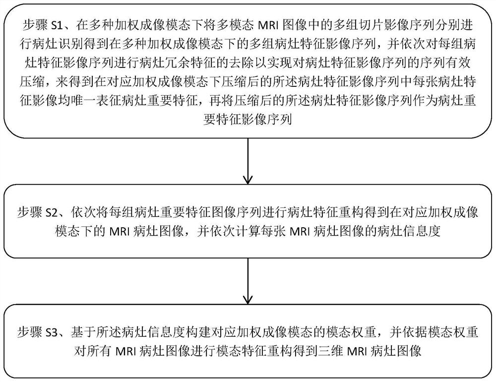
[0067] Such as figure 1 As shown, in the process of MRI imaging, by changing the influencing factors of the MR signal, different slice images can be obtained. These different slice images are called slice image sequences. For example, weighted according to the T1 value, the T1 slice image sequence can be obtained According to the T2 value weighting, the T2 slice image sequence can be obtained. One case can have multiple slice image sequences, T1 slice image se...
PUM
 Abstract
Description
Claims
Application Information