Pathological image segmentation method and device
A pathological image and image technology, applied in the field of pathological image segmentation methods and devices, can solve the problems of inability to better pay attention to the patient's condition, surgery, heavy workload for pathologists, and misdiagnosis.
- Summary
- Abstract
- Description
- Claims
- Application Information
AI Technical Summary
Problems solved by technology
Method used
Image
Examples
Embodiment 1
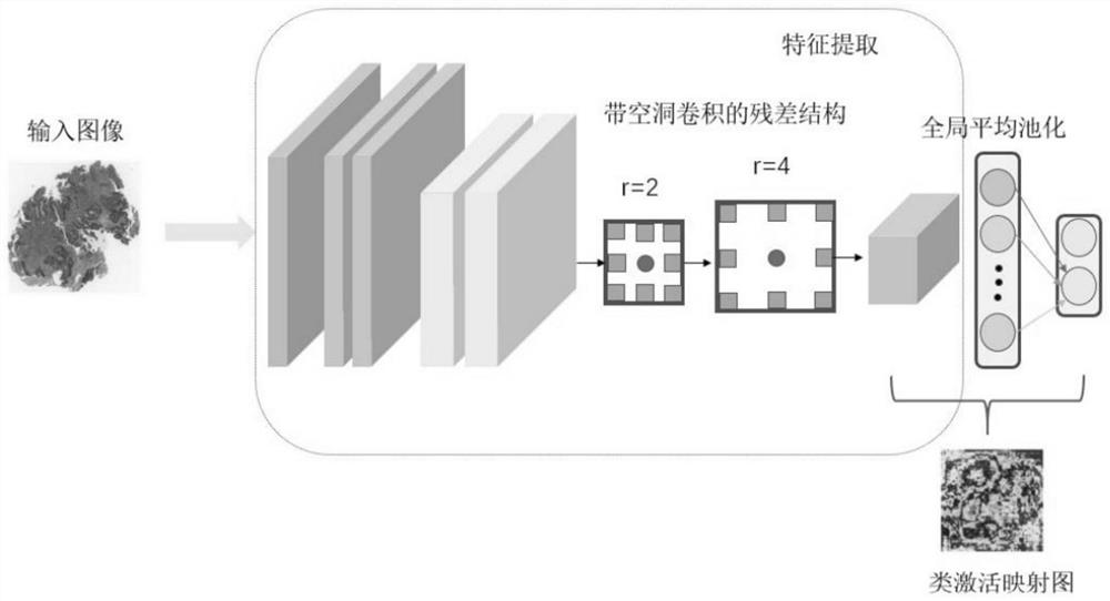
[0065] figure 1 It is a flow chart of the pathological image segmentation method provided in Embodiment 1 of the present application. This embodiment is applicable to the scene of segmenting pathological images. The method can be executed by the pathological image segmentation device provided in the embodiment of the present application. The device It can be implemented by means of software and / or hardware, and can be integrated into electronic equipment.
[0066] Such as figure 1 As shown, the segmentation method of the pathological image comprises:
[0067] S110, acquiring a pathological image of the tissue, performing preprocessing on the pathological image to obtain a preprocessed image, and using the preprocessed image as a sample and dividing it into a training set and a verification set according to a preset ratio.
[0068] Among them, in order to allow the prior classification network and the segmentation network to process various types of histopathological images, ...
Embodiment 2
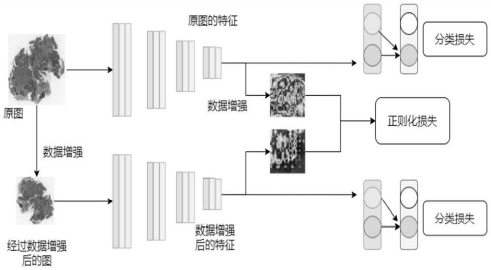
[0133] This embodiment is a preferred embodiment provided on the basis of the above-mentioned embodiments. Figure 4 is a schematic diagram of the semantic segmentation network provided in Embodiment 2 of the present application. The implementation example of the weakly supervised histopathological image segmentation method based on pseudo-label correction provided by this program includes the following steps:
[0134] Step 1: Read the histopathological image data, flip along the x-axis with a probability of 0.5, called vertical flip, flip along the y-axis with a probability of 0.5, called horizontal flip, and then perform random angles on the flipped data The rotation direction is counterclockwise, and the degree of rotation is one of the following four types: 0°, 90°, 180°, and 270°. If the degree of rotation is 0, then no rotation occurs.
[0135] The way to standardize the image is Z-Score standardization, which converts the distribution of histopathological images into a...
Embodiment 3
[0168] Figure 5 It is a structural block diagram of a pathological image segmentation device provided in Embodiment 3 of the present invention. The device can execute the pathological image segmentation method provided in any embodiment of the present invention, and has corresponding functional modules and beneficial effects for executing the method.
[0169] The device can include:
[0170] A preprocessed image acquiring unit 510, configured to acquire a pathological image of the tissue, perform preprocessing on the pathological image to obtain a preprocessed image, and divide the preprocessed image into a training set and a verification set according to a preset ratio by taking the preprocessed image as a sample;
[0171] A priori classification loss calculation unit 520, configured to input the training set sample to the prior classification network to obtain the predicted category of the sample; calculate the binary cross-entropy loss according to the predicted category a...
PUM
 Abstract
Description
Claims
Application Information
 - R&D
- Intellectual Property
- Life Sciences
- Materials
- Tech Scout
- Unparalleled Data Quality
- Higher Quality Content
- 60% Fewer Hallucinations
Browse by: Latest US Patents, China's latest patents, Technical Efficacy Thesaurus, Application Domain, Technology Topic, Popular Technical Reports.
© 2025 PatSnap. All rights reserved.Legal|Privacy policy|Modern Slavery Act Transparency Statement|Sitemap|About US| Contact US: help@patsnap.com


