Application of polymethyl methacrylate

A technology of polymethyl methacrylate and transparency, applied in the field of materials science, can solve the problems of transparency and optical imaging defects, achieve complete and convenient imaging, and achieve remarkable technological progress

Pending Publication Date: 2022-06-07
THE OBSTETRICS & GYNECOLOGY HOSPITAL OF FUDAN UNIV
View PDF0 Cites 0 Cited by
  • Summary
  • Abstract
  • Description
  • Claims
  • Application Information

AI Technical Summary

Problems solved by technology

[0007] For the above-mentioned technical problems in the prior art, the present invention provides the application of polymethyl methacrylate material in making CUBIC transparent tissue microscope imaging object holder, and described this application will solve the prior art for larger Technical problems of clearing and optical imaging of tissue samples in the light path

Method used


Image

  • Application of polymethyl methacrylate

Examples


Embodiment 1

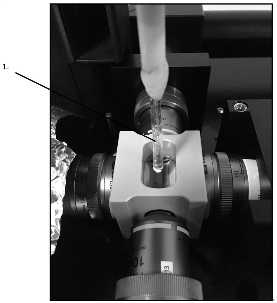
[0019] The present invention provides an application of polymethyl methacrylate material in the production of CUBIC transparent tissue microscopic imaging holder.

[0020] The present invention further provides a method for preparing a CUBIC transparent tissue microscopic imaging holder, using a polymethyl methacrylate material, the polymethyl methacrylate material is added to the blow molding device, injection device or, extrusion device molding.

[0021] Specifically, the heating temperature in the blow molding device, injection device or extrusion device is 80 ~ 300 ° C, preferably a temperature of 150 ~ 300 ° C.

[0022] Specifically, blow molding devices, injection devices or extrusion devices are prior art, which will not be repeated herein.

[0023] Specifically, the shape of the made holding vessel is tubular or sheet-shaped.

[0024] Specifically, the maximum diameter of the size of the holder made is 0.5-100 mm.

[0025]Specifically, the thickness of the made holding is ...


PUM


Abstract

The invention provides an application of a polymethyl methacrylate material in manufacturing a CUBIC transparent tissue microscopic imaging holder. The invention also provides a method for preparing the CUBIC transparent tissue microscopic imaging holder, which comprises the following steps: adding a polymethyl methacrylate material into a blow molding device, an injection device or an extrusion device for molding. The polymethyl methacrylate material can be reshaped into holding devices with various shapes and sizes after being heated, and is suitable for various biological tissue samples after CUBIC transparency, such as zebra fish, mouse embryo, brain and human tumor tissues. The holder prepared from the material disclosed by the invention can be fixedly transferred in a light sheet microscope sample bin, and a sample to be shot can be quickly and conveniently placed in the holder, so that the transparent sample can be imaged more completely and conveniently.

Description

Technical field [0001] The present invention belongs to the field of materials science, involving polymethyl methacrylate, specifically polymethyl methacrylate material in the production of CUBIC transparent tissue microscopic imaging holder application. Background [0002] Transparent and optical imaging of animal tissues is a very convenient, high-resolution way to observe the three-dimensional morphology of tissues. For these larger tissue samples, how to fix them in the optical path is a question that everyone who images transparent tissue needs to answer. There are two common types of fixation methods. [0003] The first category is to place the transparent sample directly in the sample compartment. But this poses two problems. First, because the sample is not fixed by external forces, it is easy to shift during optical imaging, which will lead to distortion of the final imaging result. Second, the light microscope cannot capture the portion of the sample that is in contact ...

Claims


Application Information

Patent Timeline
IPC IPC(8): G02B21/00G02B21/26G02B21/34
CPCG02B21/00G02B21/26G02B21/34
Inventor 徐丛剑张玲张锋
Owner THE OBSTETRICS & GYNECOLOGY HOSPITAL OF FUDAN UNIV
Why Eureka
Try Eureka