Ocular analyte sensor

An analyte and sensor technology, applied in the direction of sensors, analytical materials, material excitation analysis, etc., can solve problems such as damage

Inactive Publication Date: 2005-04-13
EYESENSE AG
View PDF39 Cites 1 Cited by
  • Summary
  • Abstract
  • Description
  • Claims
  • Application Information

AI Technical Summary

Problems solved by technology

However, this system is very invasive because it must be inserted into the bloodstream using a hypodermic needle

Method used


Image

  • Ocular analyte sensor
  • Ocular analyte sensor
  • Ocular analyte sensor

Examples


example 1

[0035] Construction of an intraocular glucose sensor

[0036] The structurally stabilizing polymer of polyethylene glycol hydrogel (PEGH, Shearwater Polymers, Inc.) was used to construct an intraocular glucose sensor. PEGH was immobilized in intraocular lenses (Alcon Laboratories, 6 mm circumference, 1 mm thickness). Chemically immobilized pendant tetramethylalkaline isothiocyanate concanavalin A (TRITC-ConA, Sigma) was included in PEGH as the acceptor half, and fluorescein isothiocyanate dextran (FITC-dextran, Sigma) was included as a competitor moiety by polymerization under UV light, as described by Ballerstadt & Schultz, Anal.Chim.Acta 345, 203-12, 1997, and Russell & Pishko Anal.Chem.71 , 3126-32, 1999 as described. Although FITC-dextran was bound to TRITC-ConA, FITC fluorescence was suppressed via fluorescence resonance energy transfer. Increasing glucose concentration releases FITC-dextran and results in fluorescence proportional to glucose concentration.

[0037] F...

example 2

[0039] Implantation of Intraocular Glucose Sensors in Life

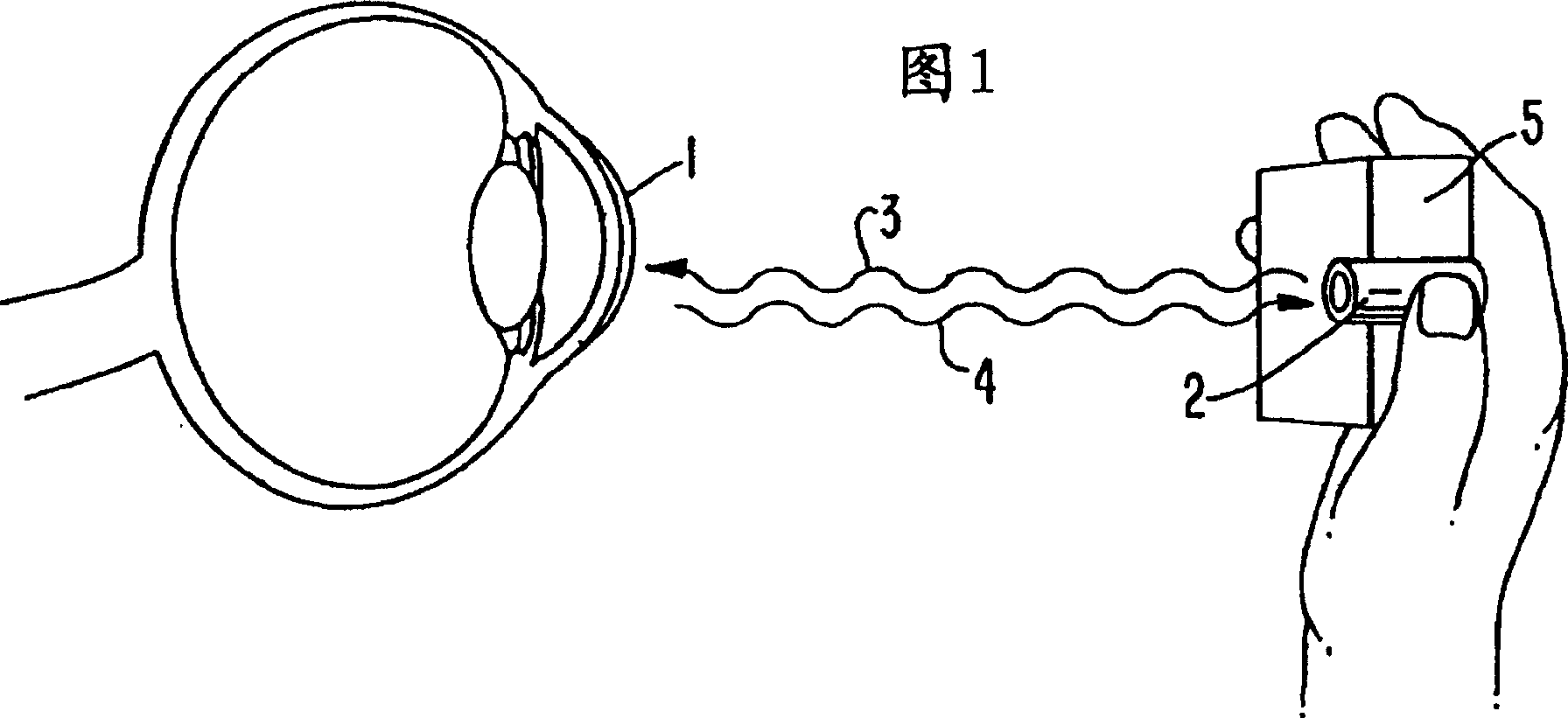
[0040] The intraocular lens glucose sensor described in Example 1 was implanted into the inner chamber of the eye of live New Zealand rabbits with a blood glucose concentration of 112 mg%. The implant is visible as a bright spot of green fluorescence (518nm) within the eye. Careful observation with the aid of a biomicroscope slit lamp indicated no toxicity, rejection, or any reaction at 6 months post-implantation.


PUM


Abstract

An ophthalmic lens comprising a receptor moiety can be used to determine the amount of an analyte in an ocular fluid. The receptor moiety can bind either a specific analyte or a detectably labeled competitor moiety. The amount of detectably labeled competitor moiety which is displaced from the receptor moiety by the analyte is measured and provides a means of determining analyte concentration in an ocular fluid, such as tears, aqueous humor, or interstitial fluid. The concentration of the analyte in the ocular fluid, in turn, inidicates the concentration of the analyte in a fluid or tissue sample of the body, such as blood or intracellular fluid.

Description

technical field [0001] An ophthalmic lens comprising a receptor half can be used to determine the amount of an analyte in eye fluid accessible to light. The receptor moiety is capable of binding a specific analyte or a detectably labeled competitor moiety. The amount of detectable competitor moiety displaced by the analyte from the receptor moiety is measured and provides a method for determining the concentration of the analyte in ocular fluids, such as tears, aqueous humor, or interstitial fluid. The analyte concentration in the eye fluid is in turn indicative of the analyte concentration in a fluid or tissue sample of the body that is not accessible, such as blood or intracellular fluid. Background technique [0002] Various nondestructive or minimally invasive methods of measuring analytes, particularly glucose, have been described. For example, US Patents 3,958,560 and 4,014,321 to March disclose a glucose sensor in which a light source on one side of the cornea is us...

Claims


Application Information

Patent Timeline
Patent Type & Authority Patents(China)
IPC IPC(8): A61B5/00G01N21/64G01N33/50G01N33/543
CPCA61B5/0002A61B5/14532A61B5/1455A61B5/411Y10S977/905A61B5/00
Inventor 韦恩·F·马克
Owner EYESENSE AG
Why Eureka
Try Eureka