

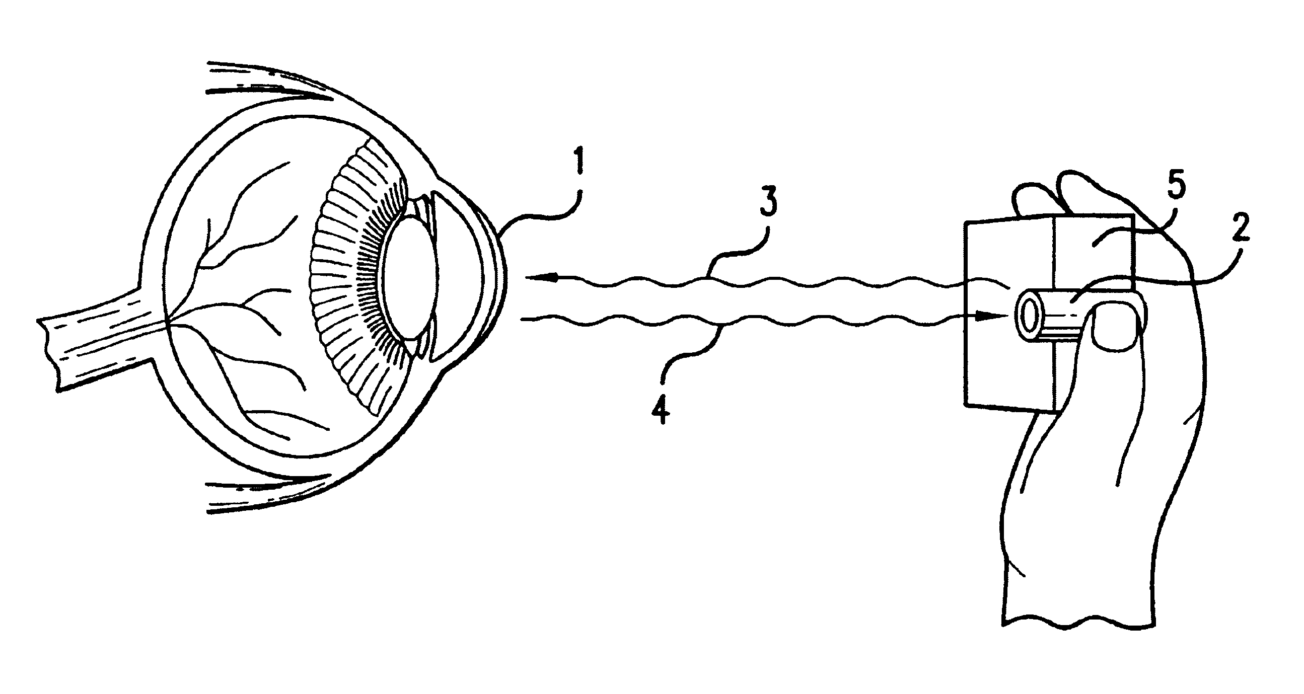
Ocular analyte sensor
- Summary
- Abstract
- Description
- Claims
- Application Information
AI Technical Summary
Benefits of technology
Problems solved by technology
Method used
Image

Examples
example 1
Construction of an Intraocular Glucose Sensor
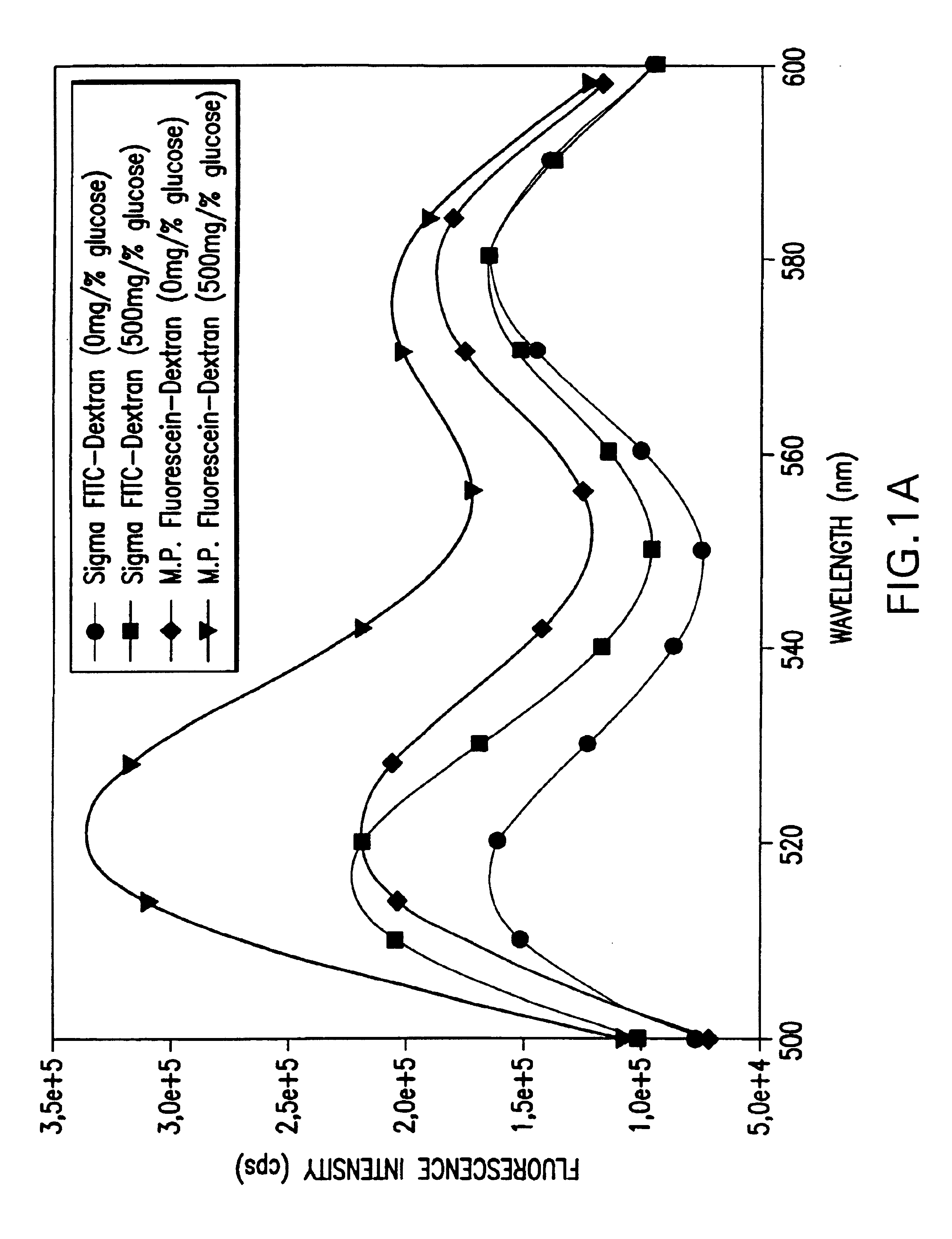
[0038]A structurally stable polymer of polyethylene glycol hydrogel (PEGH, Shearwater Polymers, Inc.) is used to construct an intraocular glucose sensor. PEGH is immobilized in an intraocular lens (Alcon Laboratories, 6 mm circumference, 1 mm thickness). Chemically immobilized pendant tetramethylrhodamine isothiocyanate concanavalin A (TRITC-ConA, Sigma) is incorporated into the PEGH as the receptor moiety and fluorescein isothiocyanate dextran (FITC-dextran, Sigma) is incorporated as the competitor moiety by polymerization under UV light, as described by Ballerstadt & Schultz, Anal. Chim. Acta 345, 203-12, 1997, and Russell & Pishko, Anal. Chem. 71, 3126-32, 1999. While the FITC-dextran is bound to the TRITC-ConA, the FITC fluorescence is quenched via a fluorescence resonance energy transfer. Increased glucose concentration frees the FITC-dextran and results in fluorescence which is proportional to glucose concentration.
[0039]FIG. 4 show...
example 2
Implantation of an Intraocular Glucose Sensor in vivo
[0040]The intraocular lens glucose sensor described in Example 1 is implanted into the anterior chamber of the eye of a living New Zealand rabbit with a blood glucose concentration of 112 mg %. The implant is visible as a bright spot of green fluorescence (518 nm) within the eye. Careful examination with a biomicroscope slit lamp shows no sign of toxicity, rejection, or any reaction 6 months after implantation.
PUM
 Abstract
Description
Claims
Application Information