i(in Vitro) induction of antigen-specific T-cells using dendritic cell-tumor cell or dendritic cell-viral cell derived immunogens

A technology of dendritic cells and tumor cells, applied in the direction of virus antigen components, cell culture active agents, fusion cells, etc.

Inactive Publication Date: 2002-06-12
UNIVERSITY OF PITTSBURGH
View PDF2 Cites 12 Cited by
  • Summary
  • Abstract
  • Description
  • Claims
  • Application Information

AI Technical Summary

Problems solved by technology

[0015] None of the above articles or patents teaches or suggests the generation of unique antigen-specific T-cells as disclosed in the present invention
In addition, none of the documents provide a significant advance of the present invention, that is, without the isolation and / or identification of specific antigens

Method used


Image

  • i(in Vitro) induction of antigen-specific T-cells using dendritic cell-tumor cell or dendritic cell-viral cell derived immunogens
  • i(in Vitro) induction of antigen-specific T-cells using dendritic cell-tumor cell or dendritic cell-viral cell derived immunogens
  • i(in Vitro) induction of antigen-specific T-cells using dendritic cell-tumor cell or dendritic cell-viral cell derived immunogens

Examples


Embodiment 1-D

[0054] Fusion of Example 1-DC and Tumor Cells

[0055] Dendritic cells were prepared from bone marrow as generally described in Celluzzi et al., J. Exp. Med. 183:283-287 (1996), in which GM-CSF was used in reference. Briefly, bone marrow cells were depleted of lymphocytes and cultured in 10% FCS containing RPMI 1640 from Irvine Scientific, Santa Ana, California at a cell density of 5×10 5 Cells / ml, using granulocyte-macrophage colony-stimulating factor (GM-CSF) from Sigma Chemical Company, St.Louis, Missouri, at a concentration of 10 3 U / ml. Loosely attached cells were collected for fusion on day 6. The concentrations of CD86 (B7.2) and MHC class II (I-A+) antigens expressed by DCs were 50-75% as measured by flow cytometry.

[0056] On day 6, DCs were fused with B16 or 3LL cells at a ratio of DCs to tumor cells of 6:1, using polyethylene glycol at 37°C. After washing by centrifugation, the fused cells were cultured overnight in RPMI1640 (10% FCS) at 37°C.

Embodiment 2

[0057] Example 2 - Preparation of co-culture of dendritic cells and tumor cells

[0058] Dendritic cells were prepared according to the method in Example 1. On day 6, dendritic cells were used to form co-cultures with B16 cells or 3LL cells. Various DCs and tumor cells were placed in test tubes to prepare various DC / tumor cell co-cultures. Centrifuge to form a cell pellet. Cell pellets were then diluted with RPMI (10% FCS) and incubated overnight at 37C in a 5% CO2 incubator. The ratio of DC:tumor cells is about 6:1. Co-cultured products were prepared to further assess whether tumor antigens were closely associated with DCs, and to assess whether soluble factors released from tumors were present on DCs.

Embodiment 3

[0059] Embodiment 3 - the effect of fusion and co-cultivation

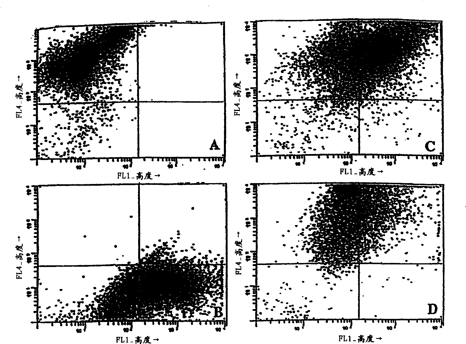
[0060] To determine the effect of fusion and co-culture, various cell types - DC, B16 and 3LL - were stained with different lipophilic fluorescent dyes prior to fusion and analyzed by flow cytometry. Tumor cells were stained with DiO and DCs were stained with DiI, both obtained from Molecular Probes, Inc., Eugene, Oregon. After extensive washing, cells were fused or co-cultured and incubated overnight at 37°C. The resulting cells were then fixed in 2% paraformaldehyde and then characterized by an Argon / HeNe duellaser on a Becton Dickinson Facstar Plus, available from Becton Dickinson Immunocytometry Systems, San Jose, CA.

[0061] For scatter characteristics of various cell types see figure 1 . Staining of individual cells showed two different patterns, with DC (DiI) shifting upwards compared to unstained controls (not shown) ( figure 1 Upper left quarter of A) or B16 tumor cells (DiO) moving to t...


PUM


Abstract

Antigen-specific T-cells prepared by culturing T-cells in formulations comprising combinations of DCs and either tumor cells or virally infected cells are disclosed. These formulations generally comprise hybridoma of at least one dendritic cell fused to either at least one tumor cell or at least one virally infected cell, or co-cultures of dendritic cells and either tumor cells or virally infected cells. The resulting T-cells can then be used in immunotherapy methods through adoptive transfer of autologous antigen-specific T-cells into patients using well-established techniques, as agents to identify tumor antigens, and to establish animal models.

Description

[0001] Cross-references to related applications [0002] This application is a continuation of U.S. Serial Application No. 09 / 030,985, filed February 26, 1998, which claims priority from U.S. Serial Application No. 60 / 039,472, filed February 27, 1997 . technical field [0003] The present invention generally relates to antigen-specific T-cells and methods of making and using them. More particularly, the present invention relates to antigen-specific T-cells produced by co-culturing with immunogens obtained from hybridomas or dendritic cells containing dendritic cells and tumor cells and tumor cell co-culture products. Alternatively, virus-infected cells can be substituted for tumor cells in these hybridomas and co-cultures. The use of these T-cells as prophylactic and therapeutic agents against tumors and viral infections is also the subject of the present invention. Background technique [0004] T-cell...

Claims


Application Information

Patent Timeline
Patent Type & Authority Applications(China)
IPC IPC(8): C12N15/02A61K35/14A61K39/00A61P31/12A61P35/00A61P37/02C12N5/0783C12N5/10C12N5/16C12N15/09C12Q1/02C12Q1/68
CPCC12N2502/30C12N2501/23C12N5/0636C12N5/16C12N2501/22A61K2039/5158C12N2502/99A61P31/12A61P35/00A61P37/02
Inventor 小L·D·法罗W·斯托尔库斯
Owner UNIVERSITY OF PITTSBURGH
Why Eureka
Try Eureka