Recombinant human antibody against HBsAg and its preparation method

A surface antigen, human-derived technology, applied in recombinant DNA technology, anti-animal/human immunoglobulin, using vectors to introduce foreign genetic material, etc., can solve the problems of difficult renaturation and low expression

Inactive Publication Date: 2005-01-26
余宙耀 +2
0 Cites 4 Cited by

AI-Extracted Technical Summary

Problems solved by technology

[0004] For the research of genetically engineered human hepatitis B surface antibody, phage display library technology is used at home and abroad to obtain its Fab segment (fragment of antigen binding) gene, and then expres...

Abstract

The present invention discloses a recombinant human antibody against HBsAg single-chain antibody used as human hepatitis B surface antibody and its preparation method. The Fab segment gene for cloning the human HBsAg antibody by bacterial virus display technology is expressed in bacillus coli; The HBsAg single-chain antibody gene can be obtained by superposing PCR and the gene is inserted into a pronucleus cytoplasm expression carrier, the high efficiency expression can be obtained in the bacillus coli, then the recombinant single-chain antibody is purified and recovered to original property; the recombinant human antibody against HBsAg single-chain antibody used as human hepatitis B surface antibody has an activity combined with the HBsAg, and the primary structure of its protein is quite correct by measuring mass spectra. The single-chain antibody constructed in the invention can replace the blood source antibody, and can be used for passive immunity of hepatitis B and can be developed for guide medicine having curing function based on it.

Application Domain

Immunoglobulins against animals/humansFermentation +1

Technology Topic

AntibodyHBsAg +12

Image

  • Recombinant human antibody against HBsAg and its preparation method
  • Recombinant human antibody against HBsAg and its preparation method
  • Recombinant human antibody against HBsAg and its preparation method

Examples

  • Experimental program(1)
  • Effect test(1)

Example Embodiment

[0060] The present invention will be further described below in conjunction with the accompanying drawings and specific embodiments, but the embodiments are only used to illustrate the present invention rather than limit the present invention.
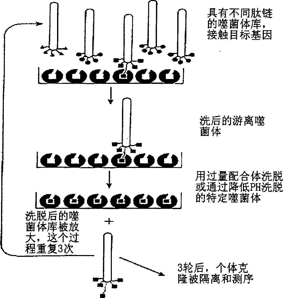
[0061] 1. Obtaining anti-HBsAg Fab antibody fragment gene by phage display technology
[0062] On the fifth day after the booster injection of hepatitis B vaccine volunteers, blood was collected, monocytes were isolated, mRNA was extracted, cDNA was synthesized by reverse transcription, and the light chain gene and heavy chain Fd of the anti-HBsAg antibody were amplified by PCR with the designed primers. The fragment genes were respectively inserted into the corresponding sites of the vector pComb3H (Carlos F.Barbas and Dennis R.Burton, 1994, Cold Spring Harbor Laboratory, Monoclonal Antibodies from combinatorial Libraries) to construct vector plasmids containing antibody light chain and heavy chain Fd segments. Transform the competent bacterium XL1-Blue with this plasmid, and the recombinant bacterium is in SB medium [Super-Broth, contains 3% Tryptone (purchased from OXOID company), 2% yeastextract (purchased from OXOID company), 1% Mops, PH7. 0] for 2 hours and then added phage VCSM13 (CarlosF.Barbas and Dennis R.Burton, 1994, Cold Spring Harbor Laboratory, Monoclonal Antibodies from combinatorial Libraries) for overnight cultivation, and the supernatant was the phage antibody library.
[0063] 1. Primer design
[0064] Heavy chain (VH) 5' primer:
[0065] VH1a CAGGTGCAGCTCGAGCAGTCTGGG
[0066] VH1f CAGGTGCAGCTGCTCGAGTCTGGG
[0067] VH2f CAGGTGCAGCTACTCGAGTCGGG
[0068] VH3aGAGGTGCAGCTCGAGGAGTCTGGG
[0069] VH4f CAGGTGCAGCTGCTCGAGTCGGG
[0070] VH4g CAGGTGCAGCTACTCGAGTGGGG
[0071] VH6a VAGGTACAGCTCGAGCAGTCAGG
[0072] Heavy chain 3' primer:
[0073] CG1a GCATGTACTAGTTTTGTCACAAGATTTGGG
[0074] CG2a CTCGACACTAGTTTTGCGTCCAACTGTCTT
[0075] CG3a TGTGTGACTAGTGTCACCAAGTGGGGTTTT
[0076] CG4a GCATGAACTAGTTGGGGGACCATATTTGGA
[0077] K chain 5' primer;
[0078] V1a GACATCGAGCTCACCCAGTCTCCA
[0079] V2a GATATTGAGCTCACTCAGTCTCCA
[0080] V3a GAAATTGAGCTGACGCAGTCTCCA
[0081] K chain 3' primer:
[0082] GCGCCGTCTAGAACTAACACTCTCCCCTGTTGAAGCTCTTTGTGACGGGCGATC
[0083] TCAG
[0084] 2. PCR amplification
[0085] Take 1ul of the above-mentioned reaction product as a template, 10 times of PCR buffer (500mM KCl, 100mM Tris.ClPH8.3, 20mM MgCl 2 ), 1.5ul of 0.1mM dNTP, 1.2ul of 20uM different combinations of 3', 5' end primers, 0.5ul of Vent DNA polymerase, 12.75ul of double distilled water. The reaction was repeated for 30 cycles (94°C*45 seconds, 50°C*60 seconds, 72°C*120 seconds), and the reaction product was recovered by 1.2% agarose electrophoresis.
[0086] 3. Screening of phage antibodies
[0087]Add 50ul of the complete set of antibody library solution to each well of the microwell plate coated with HBsAg, incubate at 37°C for 2 hours, discard the supernatant, and use an appropriate amount of 0.5% Tween20 TBS (containing 50mmol/L Tris Base, 150mmol/L NaCl) washed several times. Add 50ul of elution buffer to each well, suck out the liquid in the well, let it stand at room temperature for 10min, and adjust the pH to neutral with an appropriate amount of 2mol/L Tris. Infect 2ml of freshly prepared XL1-blue with the eluent, culture for 6-8 hours, add helper phage VCSM13 to continue culturing overnight, and re-obtain phages, repeat the above steps, collect infected bacteria after 3 rounds of screening, and extract double-stranded phage DNA ( containing Fab antibody gene).
[0088] 4. Sequence analysis and homology comparison of positive clones
[0089] The Fab-positive clones obtained from the screening against HBsAg are the clones containing the Fab antibody gene. Insert the light and heavy chain gene fragments of the antibody into the sequencing vector for sequencing identification, see the results figure 2 , for homology comparison see image 3.
[0090] 2. Cloning of genes and construction of prokaryotic expression vectors
[0091] 1. Cloning of anti-HBsAg single chain antibody gene
[0092] Primers were designed as:
[0093] Upstream primer P1: 5′-G AAT TCA TGC AGG TGC AGC TGG TGG AGT CT-3′
[0094] Downstream primer P2: 5′-GCA AGC TTT TAT CGA TTG ATT TCC ACC TTG GT-3′
[0095] GAATTCATGCAGGTGCAGCTGGTGGAGTCTGGGGGAGGCGTGGTCCAGCCTGG
[0096] GAGGTCCCTGAGACTCTCCTGTGCAGCCTCTGGATTCACCTTCAGTAGACATGG
[0097] CATGCACTGGGTCCGCCAGGCTCCAGGCAAGGGGCTGGAGTGGGTGGCAGTTA
[0098] TATCATATGATGGAAGTAATAAATACTATGCAGACTCCGTGAAGGGCCGATTC
[0099] ACCATCTCCAGAGACAATTCCAAGAACACGCTGTATCTGCAAATGAACAGCCT
[0100] GAGAGCTGAGGACACGGCTGTGTATTACTGTGCGAAAGCTGGGGGGGCAGCA
[0101] GCTGGTACGGGGACGGCCTACTTTGACTACTGGGGCCAGGGAACCCTGGTCAC
[0102] CGTCTCCTCAGGTGGCGGAGGGAGCGGTGGAGGGGGCAGTGGCGGAGGAGGT
[0103] AGCGACATTGTGTTGACCCAGTCTCCAGGCACCCTGTCTTTGTCTTCAGGGGAA
[0104] GGAGCCACCCNTTCCTGCAGGGCCAGTCAGAGTGTTAGCAACGGCCAATTAAC
[0105] CTGGTACCAGCAGAAGCCTGGCCAGGCTCCCAGGCTCCTCATCTATGGTGCAT
[0106] CCAGCAGGGCCACTGGCATCCCAGACAGGTTCAGTGGCAGTGGGTCTGGGACA
[0107] GACTTCACTTCTCACCATCAGCAGACTGGAGCCTGAAGATTTTGCAGTGTATTAC
[0108] TGTCACCAGTATGATGGCTCCCCGGAGACGTTCGGCCAAGGGACCAAGGTGGA
[0109] AATCAATCGATAAAAGCTT
[0110] 2. Construction of expression plasmid
[0111] Primers were designed as:
[0112] Upstream primer P1: 5′-TTG GAT CCC AGG TGC AGC TGG TGG AGT CT-3′
[0113] Downstream primer P2: 5′-GCA AGC TTT TAT CGA TTG ATT TCC ACC TTG GT-3′
[0114] Using pGEM3zf-ScFv as a template, add 5ul reaction buffer (500mM KCl, 100mM Tris.Cl pH8.3, 20mM MgCl 2 ), 1.5ul of 0.1mol/L dNTPs, 1ul of 20uM P1 and P2 primers, 0.5ul of Vent DNA polymerase, and add double distilled water to 50ul. The reaction was performed for 30 cycles (95°C*45 seconds, 60°C*60 seconds, 72°C*120 seconds), and the reaction product was recovered by 1.2% agarose electrophoresis. The PCR product was recovered and digested, and inserted into the pQE40 vector (purchased from QIAGEN, see QIAexpress Detection and Assay Handbook for details) under the same conditions to obtain the expression plasmid pQE-ScFv; the sequence determination results showed that the recombinant ScFv ORF (open reading frame) sequence was consistent with the expected results.
[0115] 3. Prokaryotic expression and production of human anti-HBsAg single chain antibody
[0116] 1. Induced expression of recombinant single-chain antibody
[0117] Recombinant plasmid pQE-ScFv transforms and expresses host bacterium M15[pREP4] (purchased from QIAGEN Company, see QIAexpress® Detection and Assay Handbook for details), selects a single clone and inoculates 2ml containing ampicillin (Amp) 100mg/L, kanamycin (kanamycin) ) 25mg/L LB medium [Luria-Bertani, containing 1% Tryptone (purchased from OXOID company), 0.5% yeast extract (purchased from OXOID company), 1% Nacl], cultured overnight at 37°C with shaking, and pressed 5 in the next morning % inoculum amount was transferred to LB medium containing the same antibiotic, shaken vigorously at 37°C for about 3 hours until the OD value was 0.8-1.0, and added isopropyl-b-thiogalactopyranoside (IPTG) (purchased from Huamei Biotechnology Co., Ltd. engineering company) to a final concentration of 1 mmol/L, continue to cultivate for 5 hours, collect the thalli by centrifugation, and identify the expression level of the target protein by electrophoresis, and the expression level of the target protein is 30%.
[0118] 2. Lysis of bacterial cells
[0119] Harvest the cells by centrifugation at 6000rpm, 20min, 4°C, add lysis buffer TE (10mM Tris, 1mM EDTA, pH8.0) according to 10% of the weight of the wet bacteria, wash once, discard the supernatant, add the same volume of TE, and add Lysozyme (purchased from Huamei Bioengineering Co., Ltd.) at 3‰ of the weight of the cells was mixed and stored overnight at -20°C. The next day, thaw in the refrigerator at low temperature, put in an ice bath at 400W, 5 seconds, stop for 10 seconds, sonicate a total of 10 times, 10,000rpm, 20min, and centrifuge at 4°C to collect inclusion bodies.
[0120] 3. Washing of inclusion bodies
[0121] (1) Resuspend inclusion bodies in TE, wash once at 12,000rpm, 20min, 4°C, discard the supernatant, and the remaining part is inclusion bodies;
[0122] (2) Resuspend inclusion bodies in TE containing 1% Trition X-100, wash once at 12,000rpm, 20min, 4°C, discard the supernatant, and the rest is inclusion bodies;
[0123] (3) Resuspend inclusion bodies in TE containing 2M urea, wash once at 12,000rpm, 20min, 4°C, discard the supernatant, and the remaining part is inclusion bodies;
[0124] (4) Use buffer (8M urea, 0.1M Na 2 HPO 4 , 0.01M Tris, pH8.0, 10mM β-mercaptoethanol) to lyse inclusion bodies, centrifuge at 12,000rpm for 20min, 4°C, and filter the supernatant with a 0.45 filter membrane to obtain a clarified inclusion body lysate.
[0125] 4. Purification and renaturation of recombinant single-chain antibody
[0126] 1. Purification of ScFv: a two-step purification method, the first step is Ni 2+ Ionic metal chelation chromatography, the second step is ion exchange chromatography.
[0127] (1)Ni 2+ Ion metal chelation chromatography: Since the N-terminus of the ScFv expression product is fused with a 6×His tag, it can be purified with a nickel ion chelation affinity chromatography column (purchased from Pharmacia). First equilibrate the column with buffer, the buffer is 6M urea, 0.1M NaH 2 PO 4 , 0.01M Tris, pH8.0; then load the sample, wash the column with the same buffer, and then use 4M urea, 0.1M NaH 2 PO 4 , 0.01M Tris, pH6.3 buffer elution, finally with 4M urea, 0.1M NaH 2 PO 4 , 0.01M Tris, pH4.0 buffer to elute the target protein, and the purity of the target protein after purification can reach more than 90%.
[0128] (2) Ion-exchange chromatography: the sample purified in the first step is put on SP-Sepharose CL-4B column (purchased from Pharmacia Company), and finally 4M urea, 0.01M NaH 2 PO 4 , 1M NaCl pH5.5 buffer solution elution, the target peak was collected, and analyzed by gel imaging, the purity of the target protein reached more than 97%.
[0129] 2. Refolding of ScFv
[0130] Adjust the protein concentration to 0.5mg/ml, and perform renaturation by direct dilution combined with step-by-step dialysis. The steps are as follows:
[0131] buffer containing 4M urea (0.1M NaH 2 PO 4, 0.1M Tris) dialyzed for 24 hours, then dialyzed with the same buffer solution containing 2M urea for 24 hours, then dialyzed with the same buffer solution containing 1M urea for 24 hours, then dialyzed with the same buffer solution of 0.5M urea for 24 hours, and then dialyzed with the same buffer solution containing 0.5M urea for 24 hours, and then dialyzed with The same buffer solution of 0M urea was dialyzed for 24 hours;
[0132] Add 400mM arginine (purchased from Beijing Dingguo Biotechnology Development Center) and GSSG (oxidized glutathione, purchased from Huamei Bioengineering Company) 50 times the molar concentration of ScFv when dialysis to 1M urea; 5% mannitol was added as a stabilizer during 0M urea dialysis to reduce protein aggregation and precipitation.
[0133] 5. Activity analysis and structure confirmation of ScFv
[0134] 1. Competitive ELISA
[0135] Refer to the literature (Basic Immunology Experiment Guide; Immunology Teaching and Research Office, Fourth Military Medical University, 1990.12), coat 50ng of HBsAg antigen, add a fixed amount (7ng/well) of mouse anti-HBsAg monoclonal antibody, and respectively use 10ng to 80ng The ScFv was used as a competitive antibody to compete with the mouse antibody, and the goat anti-mouse-HRP was used as the enzyme-labeled antibody, and the test was performed after color development. 450/630. The results of direct ELISA showed that the purified ScFv after refolding had strong antigen-binding ability. On the basis of this, a competitive ELISA experiment was carried out, and the amount of mouse-derived anti-HBsAg monoclonal antibody added was 7ng. It can be seen that the refolded single-chain antibody can compete with the mouse-derived anti-HBsAg monoclonal antibody, thereby inhibiting the mouse-derived anti-HBsAg monoclonal antibody. The inhibitory rate of HBsAg monoclonal antibody binding to HBsAg was 40.3%.
[0136] 2. Determination of single-chain antibody affinity constant
[0137] Noncompetitive enzyme immunoassay. Coat the ELISA plate with HBsAg at four concentrations of 3mg/L, 1.5mg/L, 0.75mg/L, and 0.375mg/L; adjust the concentration of the single-chain antibody protein to 10 -7 mol/L level, doubling dilution 1:2~1:256, add to reaction wells with different coating amounts; add mouse RGS-His TM Antibody and HRP-labeled goat anti-mouse IgG were reacted, TMB was developed, and the absorbance value at 450nm/630nm was measured. Its affinity constant was determined to be 0.23×10 by non-competitive enzyme immunoassay 8 mol/L.
[0138] 3. Confirmation of molecular weight
[0139] After SDS-PAGE electrophoresis, the refolded target protein was scanned to reach a purity of 95% ScFv, dialyzed and desalted, and used a matrix-assisted laser desorption time-of-flight mass spectrometer (MALDI-TOF-MS, BRUKER company, instrument model Bruker Reflex III, Sun Yat-sen University Analysis and Testing Center) to measure the molecular weight of ScFv, the molecular weight of the sample ScFv is 27314Da; the theoretical molecular weight calculated according to the sequence is 27322.31Da.
[0140] 4. Identification of mass peptide map

PUM

no PUM

Description & Claims & Application Information

We can also present the details of the Description, Claims and Application information to help users get a comprehensive understanding of the technical details of the patent, such as background art, summary of invention, brief description of drawings, description of embodiments, and other original content. On the other hand, users can also determine the specific scope of protection of the technology through the list of claims; as well as understand the changes in the life cycle of the technology with the presentation of the patent timeline. Login to view more.
Why Eureka
Try Eureka