Cancer immunotherapy by disrupting pd-1/pd-l1 signaling

a cancer immunotherapy and signaling technology, applied in the field of cancer immunotherapy by disrupting pd1/pdl1 signaling, can solve the problem of ineffective response, and achieve the effect of reducing or suppressing signaling and durable clinical respons

Active Publication Date: 2013-11-21
BRISTOL MYERS SQUIBB CO
View PDF1 Cites 390 Cited by
  • Summary
  • Abstract
  • Description
  • Claims
  • Application Information

AI Technical Summary

Benefits of technology

[0006]The present disclosure provides a method for immunotherapy of a subject afflicted with cancer, which method comprises administering to the subject a composition comprising a therapeutically effective amount of an agent that reduces or suppresses signaling from an inhibitory immunoregulator. In preferred embodiments, the agent is an Ab. In other preferred embodiments, the inhibitory immunoregulator is a component of the PD-1 / PD-L1 signaling pathway. In further preferred embodiments, the Ab disrupts the interaction between PD-1 and PD-L1. In certain embodiments, the Ab is an anti-PD-1 Ab of the invention or an anti-PD-L1 Ab of the invention. In preferred embodiments, the anti-PD-1 Ab of the invention is nivolumab (BMS-936558) and the anti-PD-L1 Ab of the invention is BMS-936559. In certain embodiments, the subject has been pre-treated for the cancer. In other embodiments, the cancer is an advanced, metastatic and / or refractory cancer. In preferred embodiments, the administration of the antibody or antigen-binding portion to the subject thereof induces a durable clinical response in the subject.

Problems solved by technology

Although an endogenous immune response to cancer is observed in preclinical models and patients, this response is ineffective, and established cancers are viewed as “self” and tolerated by the immune system.
However, unlike the clinical development of small molecule agents that target discrete activating mutations found in select cancer populations, a particular challenge in cancer immunotherapy has been the identification of mechanism-based predictive biomarkers to enable patient selection and guide on-treatment management.

Method used


Image

  • Cancer immunotherapy by disrupting pd-1/pd-l1 signaling
  • Cancer immunotherapy by disrupting pd-1/pd-l1 signaling
  • Cancer immunotherapy by disrupting pd-1/pd-l1 signaling

Examples


example 1

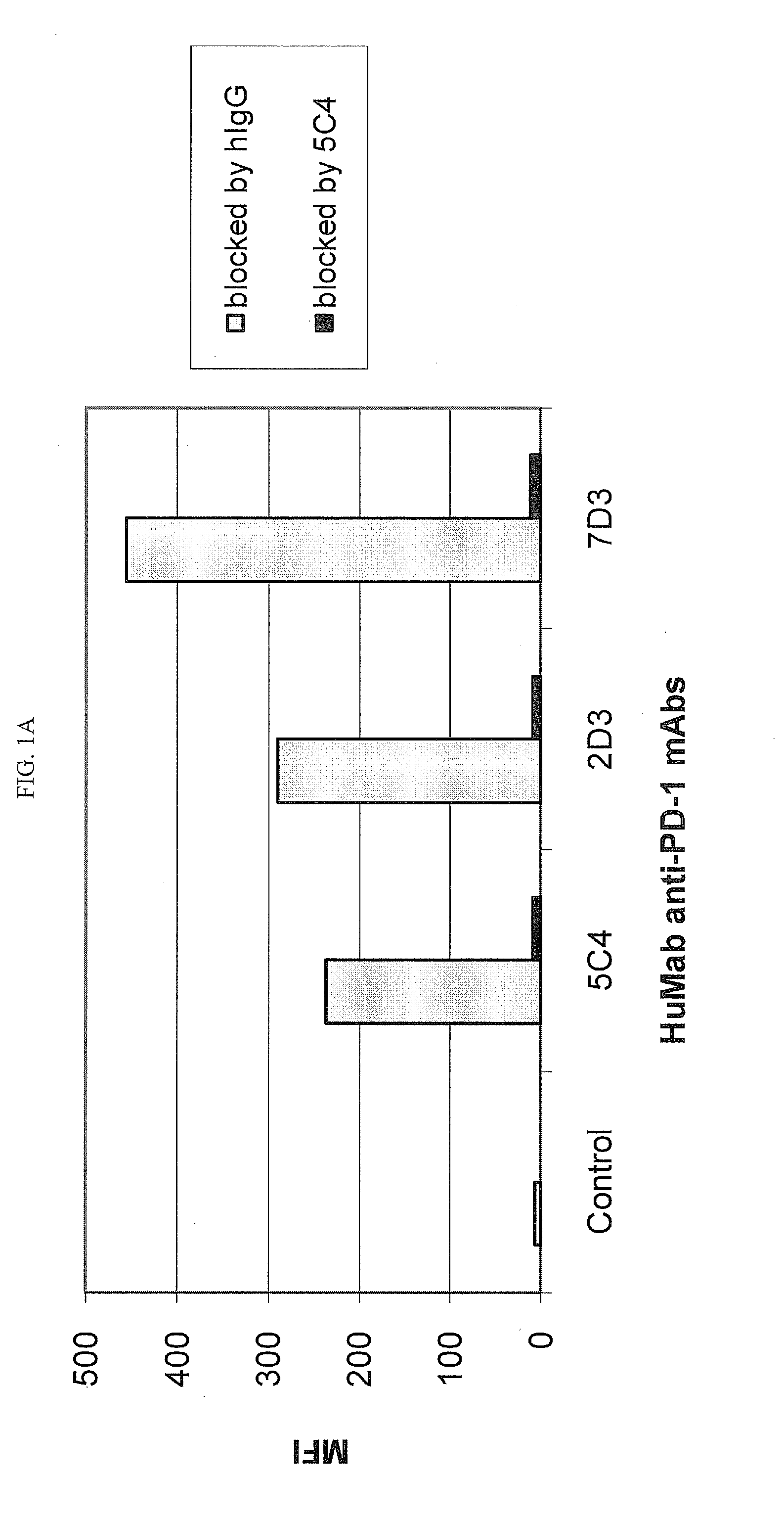
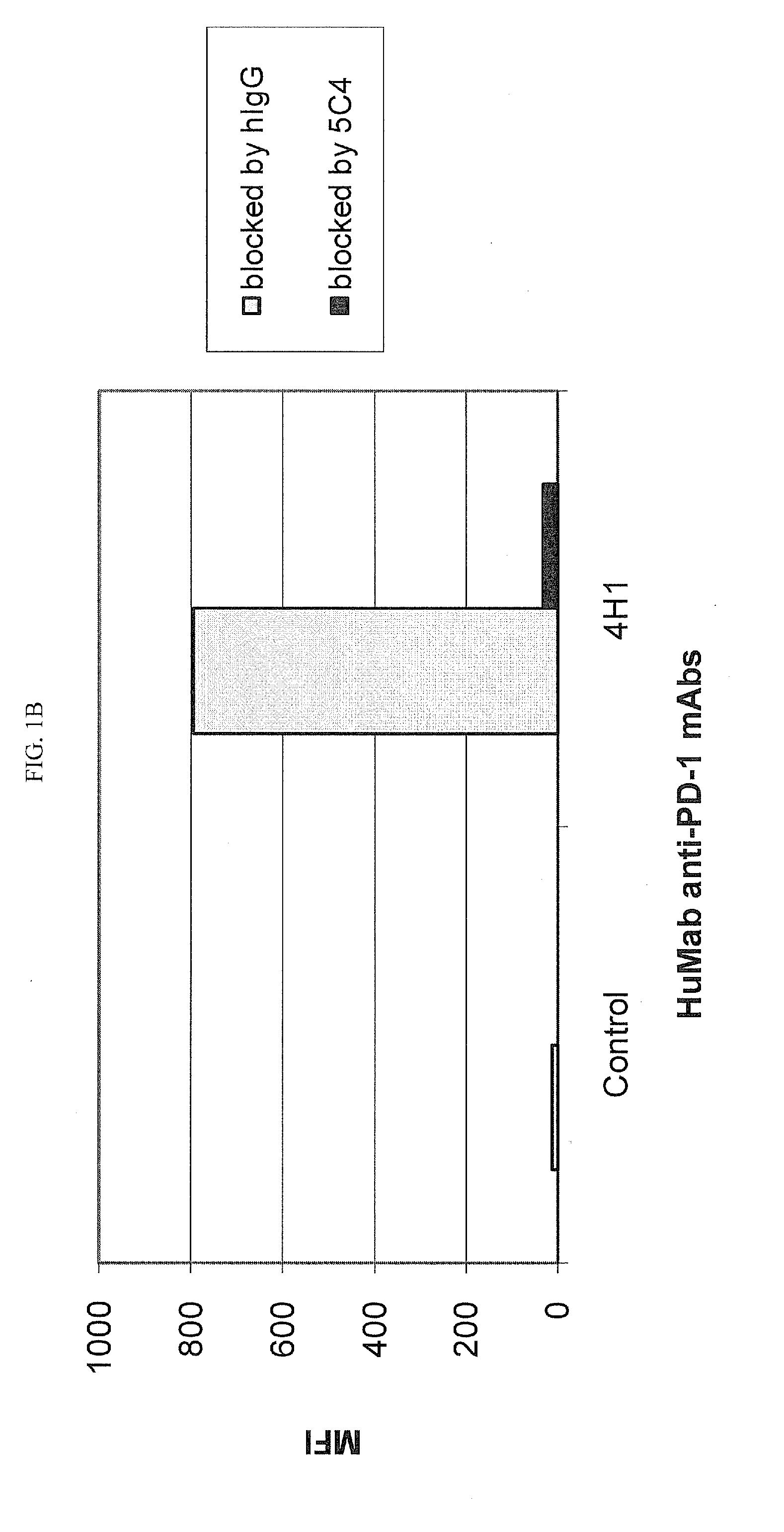
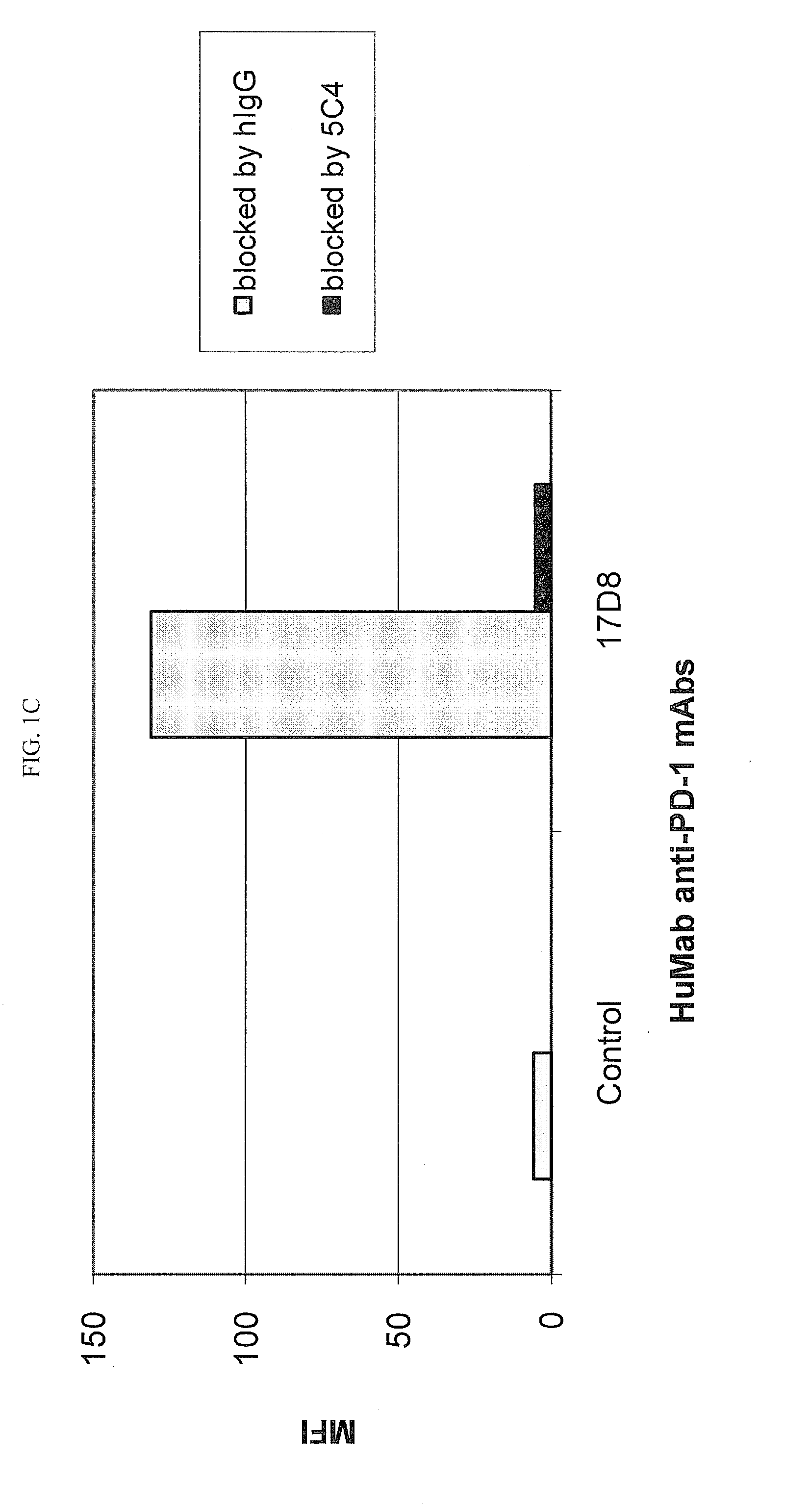
Cross-Competition Between Anti-PD-1 HuMAbs for Binding to CHO Cells Expressing Human PD-1

[0218]Chinese Hamster Ovary (CHO) cells transfected to express human PD-1 (CHO / PD-1 cells) were incubated with 10 μg / ml of Fab fragment of the anti-PD-1 HuMAb 5C4 or human IgG1 (hIgG1) isotype control Ab for 30 minutes at 4° C. before addition of anti-PD-1 HuMAbs 2D3, 7D3 or 4H1 at a concentration of 0.2 μg / ml. Binding of 4H1, 2D3 or 7D3 to CHO / PD-1 cells were detected by fluorescein isothiocyanate (FITC)-conjugated goat anti-hIgG, Fc-gamma specific Ab. In the case of cross-competition assay with 5C4 and 17D8, CHO / PD-1 cells were incubated with the whole molecule of 5C4 before addition of FITC-labeled 17D8. Binding of 2D3, 7D3, 4H1 or 17D8 to the CHO / PD-1 cells was measured by flow cytometric analysis using a FACS® calibur flow cytometer (Becton Dickinson, San Jose, Calif.).

[0219]The results are depicted in FIG. 1. The data show that the 5C4 Fab fragment substantially blocked the binding of mAbs...

example 2

Cross-Competition Between Anti-PD-L1 HuMAbs for Binding to CHO Cells Expressing Human PD-L1

[0220]CHO cells transfected to express hPD-L1 (CHO / PD-L1 cells) were incubated with 10 μg / ml of each of ten unconjugated human anti-PD-L1 mAbs (5F8, 7H1, 10H10, 1B12, 3G10, 10A5, 11E6, 12A4, 12B7, and 13G4) or human IgG1 (hIgG1) isotype control Ab for 20 min at 4° C. FITC-conjugated 10H10 (A), 3G10 (B), 10A5 (C), 11E6 (D), 12A4 (E) or 13G4 (F) was added to the cells to a final concentration of 0.09 μg / ml (B, D), 0.27 μg / ml (A, C), 0.91 μg / ml (F), or 2.73 μg / ml (E) for an additional 20 min at 4° C. without prior washout of unbound, unconjugated Ab. Different quantities of the various FITC-conjugated HuMAbs were used due to differences in binding efficiency following labeling, and the optimal amounts of these FITC-conjugated HuMAbs were previously determined by dose-titration analysis of binding to CHO / PD-L1 cells. Binding of FITC-conjugated 10H10, 3G10, 10A5, 11E6, 12A4 or 13G4 to the CHO / PD-L1...

example 3

Cross-Competition Between Anti-PD-L1 mAbs for Binding to Ovarian Carcinoma Cells Expressing Human PD-L1

[0222]Anti-PD-L1 HuMAbs 5F8, 12B7, 3G10, 1B12, 13G4, 10H10, 10A5 and 12A4, and a human IgG1 (huIgG1) isotype control Ab were serially diluted from 10 ng / ml and incubated with hPD-L1-expressing ES-2 ovarian carcinoma cells for 20 minutes at 4° C. Without washing, biotinylated-12A4 Ab was added to a final concentration of 0.4 ng / ml for an additional 20 minutes at 4° C. After washing, bound biotin-12A4 was detected using fluorescent streptavidin-PE secondary reagent and measured by flow cytometry. FIG. 3 shows the fluorescence of bound biotin-12A4 plotted against the concentration of unlabeled hPD-L1 HuMAbs. Binding of biotin-12A4 to ES-2 cells was substantially blocked by 12A4 itself and by 1B12 and 12B7, and was moderately to significantly blocked by mAbs 5F8, 10A5, 13G4 and 3G10, but was not blocked by mAb 10H10.


PUM

PropertyMeasurementUnit
thresholdaaaaaaaaaa
thresholdaaaaaaaaaa
concentrationaaaaaaaaaa

Abstract

The disclosure provides a method for immunotherapy of a subject afflicted with cancer, comprises administering to the subject a composition comprising a therapeutically effective amount of an antibody that inhibits signaling from the PD-1 / PD-L1 signaling pathway. This disclosure also provides a method for immunotherapy of a subject afflicted with cancer comprising selecting a subject that is a suitable candidate for immunotherapy based on an assessment that the proportion of cells in a test tissue sample from the subject that express PD-L1 on the cell surface exceeds a predetermined threshold level, and administering a therapeutically effective amount of an anti-PD-1 antibody to the selected subject. The invention additionally provides rabbit mAbs that bind specifically to a cell surface-expressed PD-L1 antigen in a FFPE tissue sample, and an automated IHC method for assessing cell surface expression in FFPE tissues using the provided anti-PD-L1 Abs.

Description

[0001]Throughout this application, various publications are referenced in parentheses by author name and date, or by patent No. or patent Publication No. Full citations for these publications may be found at the end of the specification immediately preceding the claims. The disclosures of these publications are hereby incorporated in their entireties by reference into this application in order to more fully describe the state of the art as known to those skilled therein as of the date of the invention described and claimed herein. However, the citation of a reference herein should not be construed as an acknowledgement that such reference is prior art to the present invention.FIELD OF THE INVENTION[0002]This invention relates to methods for immunotherapy of a cancer patient comprising administering to the patient antibodies that disrupt the PD-1 / PD-L1 signaling pathway. A biomarker may be used as part of this treatment for identifying suitable patients for immunotherapy and for pred...

Claims


Application Information

Patent Timeline
Patent Type & Authority Applications(United States)
IPC IPC(8): C07K16/28
CPCC07K16/2818C07K16/2827A61K2039/505A61K2039/507C07K2317/76A61K39/3955C07K16/18C07K16/2803G01N33/57492A61P35/00A61P35/04A61P43/00Y02A50/30G01N2333/70596G01N2800/52C07K2317/33C07K2317/92
Inventor COGSWELL, JOHNGOLDBERG, STACIE M.GUPTA, ASHOK K.JURE-KUNKEL, MARIAWANG, XI TAOWIGGINTON, JOHN M.
Owner BRISTOL MYERS SQUIBB CO
Why Eureka
Try Eureka