
Method of three dimensional display for filtered faultage radiography after reproducing three-dimensional data successfully

By using a three-dimensional model to calculate variance and two-dimensional convolution filtering in tomography images, the relationship between noise and image quality is solved, higher image details and lower dose burden are achieved, and the quality of tomography images is significantly improved. Display effect.

Inactive Publication Date: 2005-09-07
SIEMENS AG
View PDF0 Cites 7 Cited by
  • Summary
  • Abstract
  • Description
  • Claims
  • Application Information

AI Technical Summary

Problems solved by technology

[0003] The methods described above lead to Gaussian filter properties due to the "central limit value law", which for radiology generally do not correspond to the usual image impression of diagnostic images and are therefore rejected
Another problem lies in the running time of these algorithms, which is in the range of minutes for each axial slice due to the large number of iterations, thus making the method less useful clinically.

Method used


Image

  • Method of three dimensional display for filtered faultage radiography after reproducing three-dimensional data successfully
  • Method of three dimensional display for filtered faultage radiography after reproducing three-dimensional data successfully
  • Method of three dimensional display for filtered faultage radiography after reproducing three-dimensional data successfully

Examples


Embodiment Construction

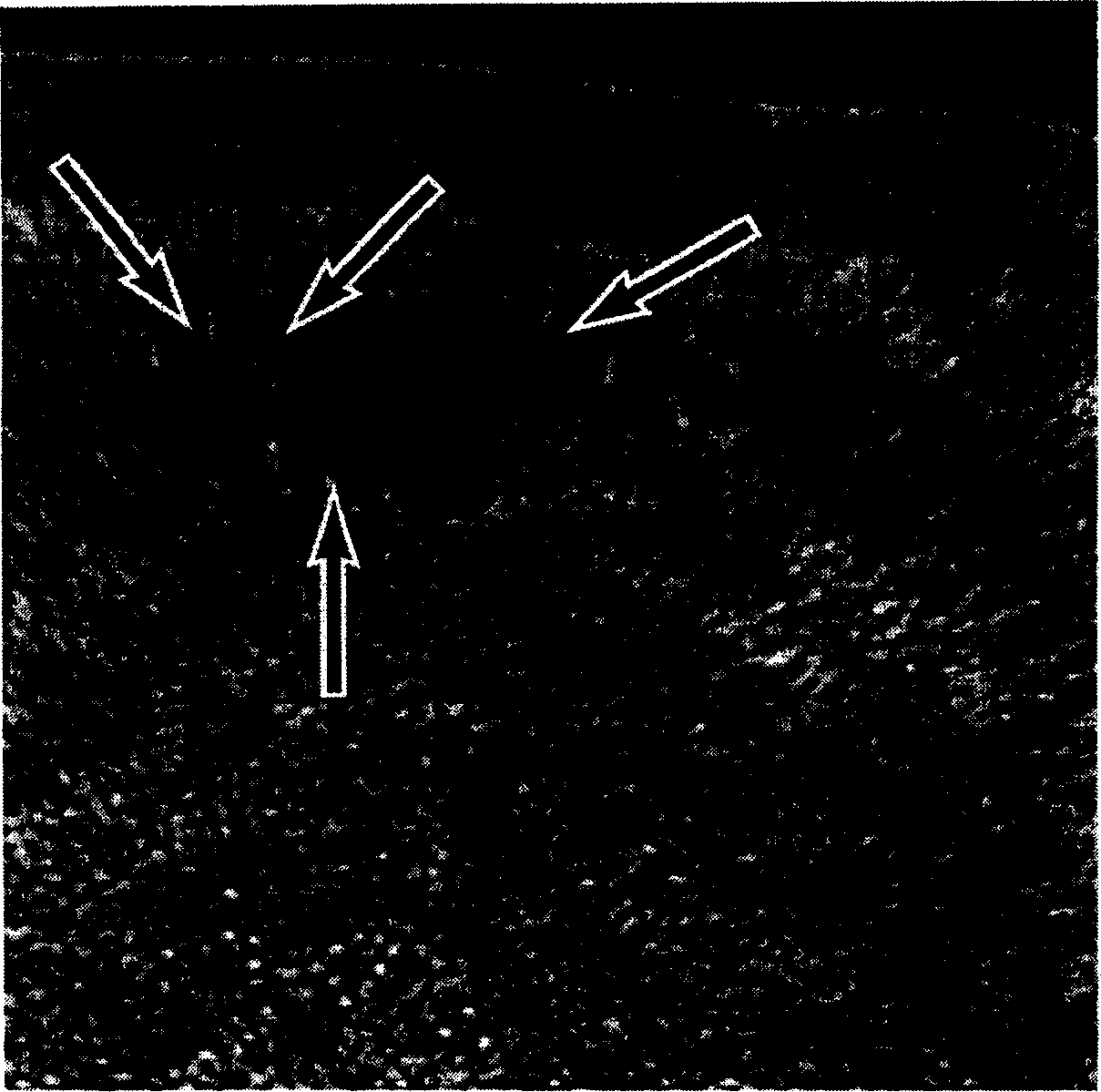
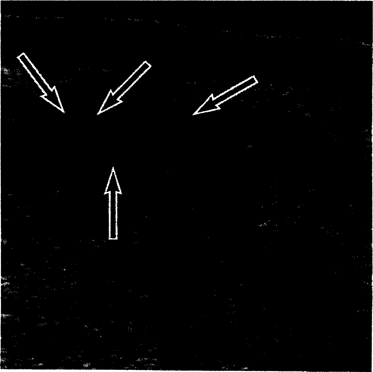
[0031] figure 1 and figure 2 The problem of linear low-pass filtering of CT photographs is shown. exist figure 1 An unfiltered CT cross-sectional image can be seen in , which is now filtered by a linear low-pass filter figure 2 middle. Although the noise is reduced as desired, the image sharpness is also reduced, small structures are lost and edges are blurred. figure 1 and figure 2 Arrows shown in indicate these problem areas.

[0032] The method according to the invention solves these problems, for example by employing the following particularly preferred method steps:

[0033] step 1:

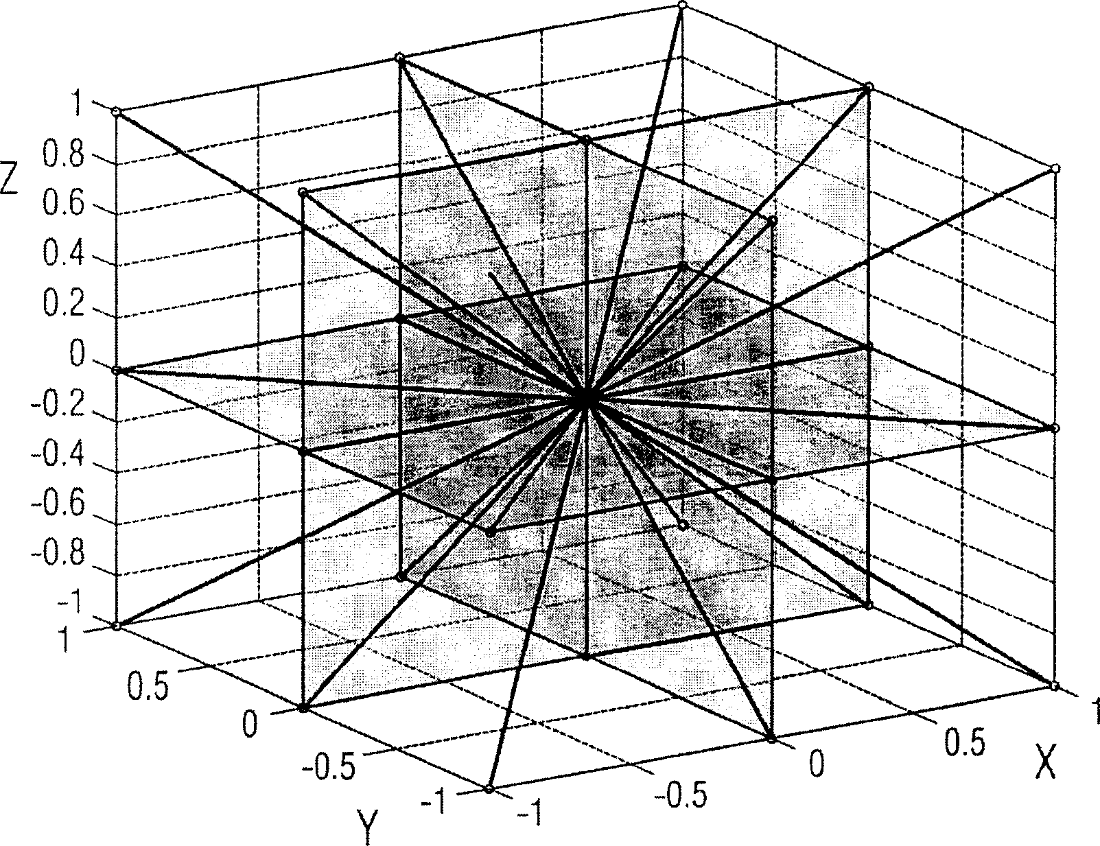
[0034] For each image voxel representing a data point in the three-dimensional space of the object under examination with coordinates x, y, z, a one-dimensional variance within a suitable radius R is calculated in a plurality of spatial directions. exist image 3 A meaningful choice of these spatial directions is shown in . 3 standard axes, 6 planar diagonals and 4 spatial di...


PUM


Abstract

A method is for filtering tomographic 3D images of an examination object. The examination object is imaged by using a volume model that divides the volume of the examination object into a multiplicity of three-dimensional image voxels with individual image values, wherein the image value of each voxel reproduces an object-specific property of the examination object in this volume. After the reconstruction of the total volume for each voxel, variances are calculated in a prescribed region or radius R in order to determine contrast jumps and their spatial orientation with their tangential planes T. The image values in the tangential plane T are filtered with a two-dimensional convolution, and subsequently the original voxel data are mixed in weighted fashion with the filtered voxel data.

Description

technical field [0001] The invention relates to a method for the three-dimensional display of a tomographic image of an examination object, preferably a filtered CT or NMR or PET image of a patient, wherein a three-dimensional model is used for displaying the examination object, which divides the volume of the examination object into multiple three-dimensional image voxels with respective image values, and the image value of each voxel gives object-specific properties of the object under examination within the volume. Background technique [0002] Methods for suppressing noise in volume data are known in principle. Noise can thus be effectively reduced by means of a linear low-pass filter, but the sharpness of the data material is reduced and thus the display quality of small structures is reduced. Therefore, this simple law can only be used to a limited extent to improve image material. Another approach is based on iterative filtering of the data material in 2D or 3D, whe...

Claims


Application Information

Patent Timeline
Patent Type & Authority Applications(China)
IPC IPC(8): G01T1/161A61B5/055A61B6/00A61B6/03G01N23/00G06K9/40G06T1/00G06T5/20G06T11/00G06T17/00G21K1/12H05G1/60
CPCG06T11/008
Inventor 丹尼尔·费希尔托马斯·弗洛尔雷纳·劳帕克
Owner SIEMENS AG
Learn More