
Method and apparatus for analyzing biological tissue images

An image and image repetition technology, applied in image enhancement, image analysis, image data processing, etc., can solve problems such as misleading, inability to correctly quantify required parameters, incorrectness, etc.

Inactive Publication Date: 2006-03-01
HUMANITAS MIRASOLE SPA
View PDF0 Cites 0 Cited by
  • Summary
  • Abstract
  • Description
  • Claims
  • Application Information

AI Technical Summary

Problems solved by technology

In this case, the known devices are not able to correctly quantify the required parameters, such as volume, and thus the results of the analysis may be incorrect or even misleading

Method used


Image

  • Method and apparatus for analyzing biological tissue images
  • Method and apparatus for analyzing biological tissue images
  • Method and apparatus for analyzing biological tissue images

Examples


Embodiment Construction

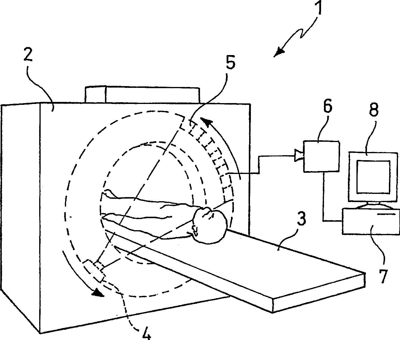
[0013] The example to be described hereinafter relates to a system 1 for acquiring and processing images comprising a conventional CAT scanner 2 with a motorized bed 3 able to pass over the CAT scanner.

[0014] The CAT scanner 2 is provided with an X-ray tube 4 and a detector box 5 positioned diametrically relative to the X-ray tube. The X-ray tube 4 and the detector box 5 can be rotated synchronously around said bed 3 in which the patient lies during the analysis.

[0015] The electronic image acquisition device 6 is operatively connected to the detector box 5 . The electronic image capture device 6 is then operatively connected to the processing system 7 . The processing system 7 may be implemented using a personal computer (PC), which includes a bus connecting processing means such as a central processing unit (CPU) with storage means including, for example, RAM working memory, read-only memory (ROM) - its Includes the basic program for booting the computer—a magnetic ha...


PUM


Abstract

The present invention relates to a method and an apparatus for processing images of biological tissues, in particular of human or animal origin. The metric quantification of a biological body part or tissue or of an abnormal material spot or aggregate contained therein is also performed. The invention is applied in particular to the Computed Axial Tomography technique. In particular, the present invention comprises the following steps: la) dividing the image into boxes of different size iteratively; (2a) calculating for each quadrant at each division scale the relative dispersion (RD) obtained as the Standard Deviation divided by the mean value of the pixels, in order to associate to each quadrant a set of values of RD; (3a) generating a homogeneity map as a grey scale image, each point's brightness being given by the mean of the set of values of RD for each quadrant, wherein the image's regions having higher brightness correspond to homogeneous regions.

Description

technical field [0001] The invention relates to a method and a device for processing images of biological tissues, especially human or animal organs. Quantification of measurements of biological body parts or tissues or spots or aggregates of abnormal material contained therein can also be carried out using the method according to the invention. [0002] The method according to the invention has particular application in computer-aided tomography. Background technique [0003] With regard to the term "abnormal speck or aggregate of material", it refers to a speck or aggregate of material morphologically associated with a pathological state or a state giving rise to a pre- or post-pathological situation. Examples of spots or aggregates of abnormal material may be tumors, atherosclerotic plaques, edemas, hematomas, acute or chronic inflammatory lesions, scars, and collagen diseases. [0004] When pathological diagnosis requires observation of body parts or organs, such obser...

Claims


Application Information

Patent Timeline
Patent Type & Authority Applications(China)
IPC IPC(8): G06T5/00G06T7/00G06T7/60
CPCG06T2207/10081G06T7/602G06T7/0081G06T2207/30096G06T7/0012G06T7/11G06T7/62
Inventor 尼古拉·迪奥瓜尔迪法比奥·格里齐卡洛·鲁索芭芭拉·弗兰切斯基尼皮尔卡洛·穆齐奥
Owner HUMANITAS MIRASOLE SPA
Learn More