Ultrasound system for internal imaging including control mechanism in a handle

An ultrasound system and handle technology, applied in the field of medical ultrasound systems, can solve the problems of prolonging the inspection time, the controller cannot be adjusted by the controller, and the sensor movement.

Inactive Publication Date: 2006-07-12
KONINKLIJKE PHILIPS ELECTRONICS NV
View PDF0 Cites 12 Cited by
  • Summary
  • Abstract
  • Description
  • Claims
  • Application Information

AI Technical Summary

Problems solved by technology

This causes sensor movement, requiring repositioning of the sensor, thus prolonging the inspection time
[0007] Another disadvantage of the pro

Method used


Image

  • Ultrasound system for internal imaging including control mechanism in a handle
  • Ultrasound system for internal imaging including control mechanism in a handle
  • Ultrasound system for internal imaging including control mechanism in a handle

Examples


Embodiment Construction

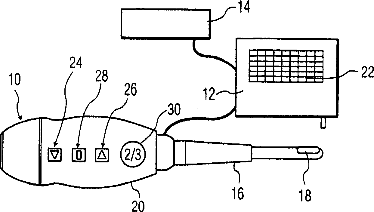
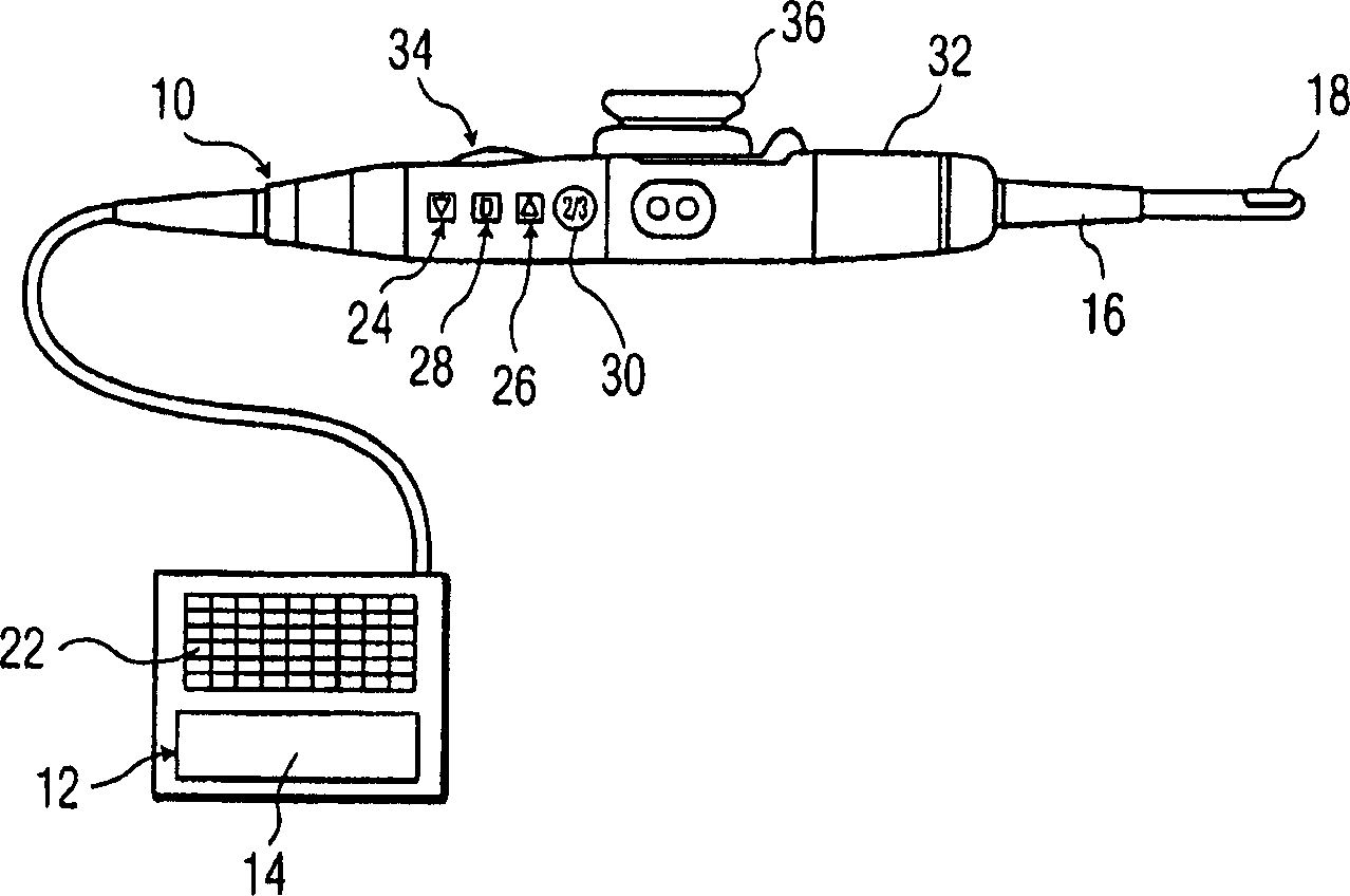
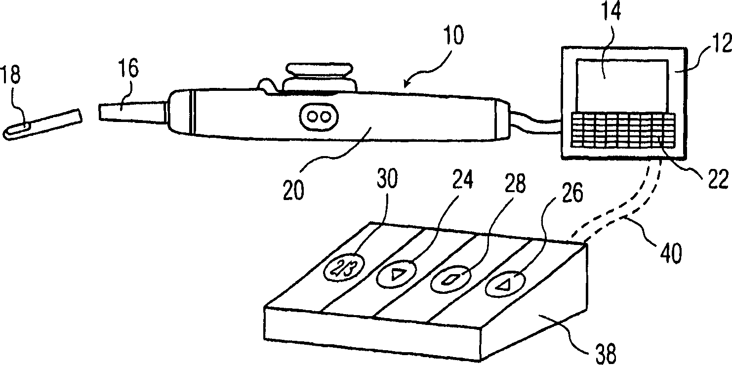
[0025] Referring to the drawings, wherein like reference numerals indicate the same or similar elements, figure 1 An ultrasound imaging system is shown comprising a transthoracic probe 10, a control unit 12 and a display device 14, such as a monitor. The probe 10 includes a handle 20 connected to the control unit 12 by a cable and an ultrasound transducer 18 arranged to be connected to the handle 20 (ie, disposed within the handle 20) to obtain images of the patient's internal structures. The control unit 12 and the display device 14 can be integrated, ie formed as a common unit.

[0026] A control panel 22 including function-specific controls is provided in connection with the control unit 12 . The function-specific controls include controls for switching the mode of operation of the probe 10 and controls for optimizing the image captured by the sensor 18 and displayed on the display device 14, among other known controls for use with the probe 10. device. The control unit ...


PUM


Abstract

Ultrasound systems for imaging internal structure of a patient including a probe ( 10 ) having a handle ( 20,32 ) and a transducer ( 18 ) arranged in connection with the handle ( 20,32 ) for obtaining images, either in the handle as in a transthoracic probe ( 10 ) or in a probe shaft ( 16 ) coupled to the handle as in a TEE probe ( 10 ). Controls on the handle enable a switch in the operation mode of the probe ( 10 ) and/or optimization of images obtained by the transducer ( 18 ). A display device ( 14 ) coupled to the probe ( 10 ) displays control menus and optional obtained images. A control unit ( 12 ) controls the probe ( 10 ) and display device ( 14 ) based on control activation. A change in control menu displayed by the display device ( 14 ), a switch in the mode of operation of the probe ( 10 ) and/or adjustment of images obtained by probe ( 10 ) is effected based on activation of handle control(s) ( 24,26,28 ). A foot pedal ( 38 ) can be provided including same controls ( 24,26,28 ) as the handle ( 20,32 ).

Description

technical field [0001] The present invention relates generally to medical ultrasound systems for internal imaging, and more particularly to medical ultrasound systems including controls within a handle that is manipulated during an examination. Background technique [0002] Various medical probes that include sensors are used to obtain images of internal organs and tissues. For example, a transesophageal echocardiography (TEE) probe includes a transducer designed to be inserted into a patient's mouth and positioned within the patient's esophagus, thereby imaging the heart and other visceral structures in the direction of view of the transducer. Likewise, transthoracic sensors are designed to be positioned within the patient's chest such that the heart and other internal structures imaged within the chest cavity are in the sensor's viewing direction. [0003] The TEE probe comprises a sensor disposed within a probe shaft adapted to be inserted into the patient, a "middle han...

Claims


Application Information

Patent Timeline
IPC IPC(8): A61B8/00A61B8/12
CPCA61B8/465A61B8/483A61B8/467A61B8/12A61B8/461
Inventor J·拜伦
Owner KONINKLIJKE PHILIPS ELECTRONICS NV
Why Eureka
Try Eureka