Method of inducing differentiation of human amnion mesenchyme stem cell to nerve cell

A technology of mesenchymal stem cells and nerve cells, applied in the field of inducing human amniotic mesenchymal stem cells to differentiate into nerve cells

Inactive Publication Date: 2006-08-02
SHENZHEN BEIKE BIOTECH
View PDF0 Cites 5 Cited by
  • Summary
  • Abstract
  • Description
  • Claims
  • Application Information

AI Technical Summary

Problems solved by technology

[0004] The first purpose of the present invention is to provide a method for inducing human amniotic mesenchymal stem cells to differentiate into nerve cells in view of the current situation that there is no method for inducing differentiation of amniotic mesenchymal stem cells into nerve cells

Method used


Image

  • Method of inducing differentiation of human amnion mesenchyme stem cell to nerve cell
  • Method of inducing differentiation of human amnion mesenchyme stem cell to nerve cell
  • Method of inducing differentiation of human amnion mesenchyme stem cell to nerve cell

Examples


Embodiment 1

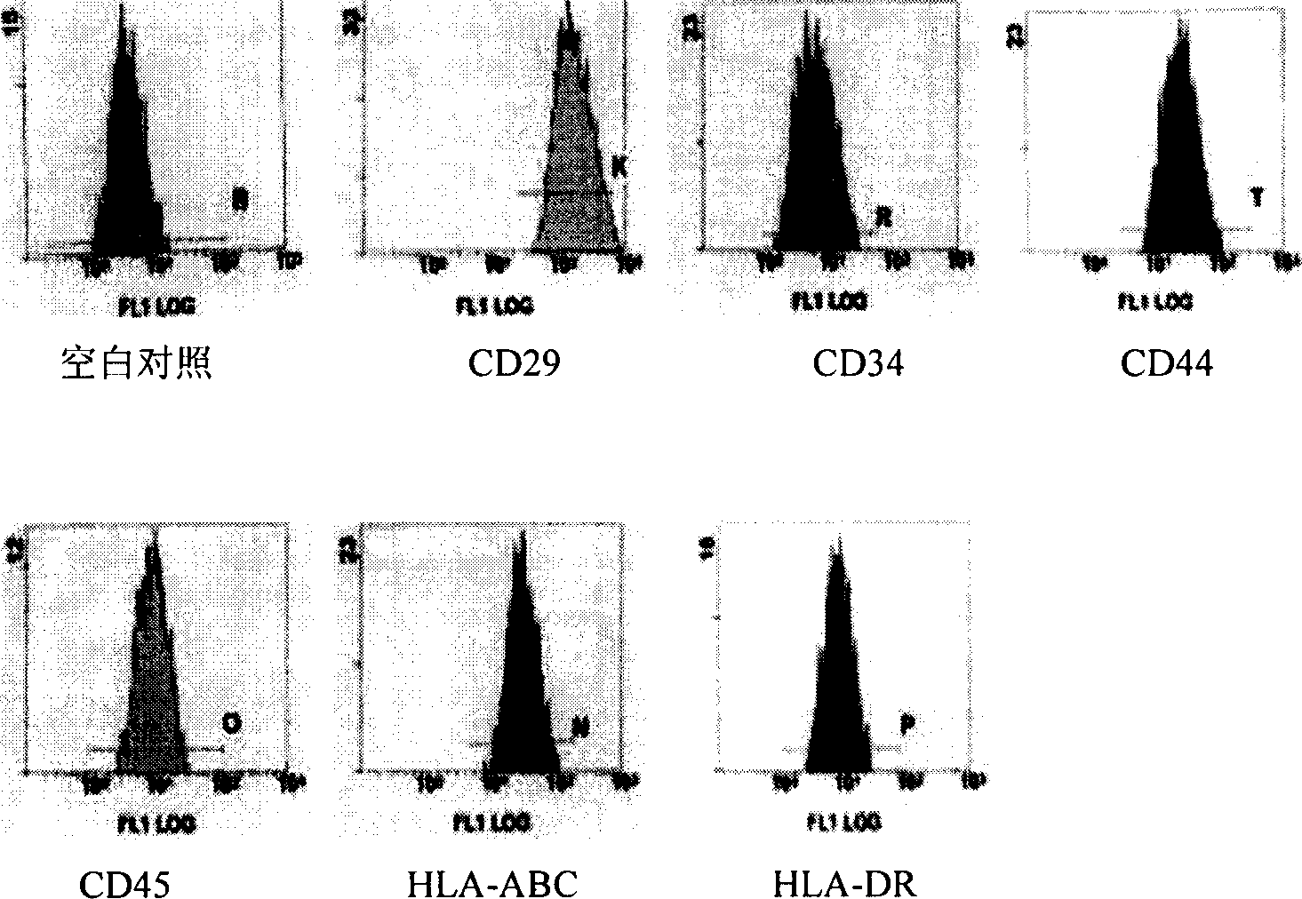
[0024] 1. Isolation, culture, expansion and purification of human amniotic membrane mesenchymal stem cells

[0025] 1. Isolation of human amniotic membrane mesenchymal cells:

[0026] Under aseptic conditions, take the human placenta of a normal term cesarean section fetus, bluntly separate the amniotic membrane from the umbilical cord of the placenta, rinse thoroughly with phosphate buffered saline (PBS), and cut the amniotic membrane into 1.0cm×1.0cm sheets, Add 2ml of 0.25% trypsin to each gram of tissue, digest it for 30 minutes at room temperature for 3 times, then stop trypsin with DMEM / F12 culture medium containing 10% fetal bovine serum (FBS) by volume. To remove epithelial cells as much as possible. Then cut the amniotic membrane as much as possible, and add V containing 1.0g / L collagenase and 0.10g / L deoxyribonuclease per gram of tissue DMEM : V F12 =1:1 DMEM / F12 culture medium 2ml, digested at 37°C for 60 minutes to obtain a cell suspension. Then filter the digested cel...

Embodiment 2

[0039] Example 2: Application of composition containing HADMSCs in the treatment of neurological diseases

[0040] HADMSCs can differentiate into nerve cells, opening up a new cell source for nerve transplantation. The present invention establishes an animal model to further confirm the above results.

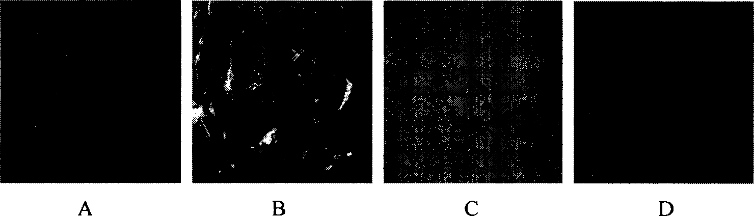
[0041] 1. Animal grouping and model making:

[0042] There are 80 Wistar rats, male or female, randomly divided into 4 groups: Group A is the injury group (Sham), 20 rats, only the craniotomy, but not the brain tissue, and the transplantation of cells, served as the negative control group. Group B was a sham transplant group (TBI+NS), with 20 rats. One day after craniotomy and drilling to strike the brain tissue, 10μl of normal saline was injected as an intervention control. Group C is the transplantation group (TBI+HADMSCs), with 20 rats. One day after craniotomy and drilling to strike the brain tissue, 10μl of normal saline containing HADMSCs was injected as the HADMSCs transplan...

Embodiment 3

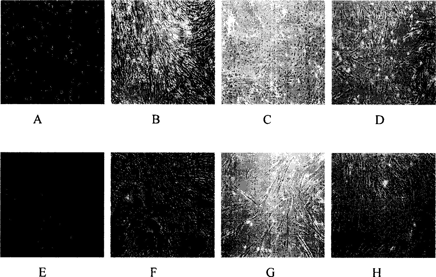
[0048] Example 3: Application of fibrin glue as a scaffold material of HADMSCs in tissue engineering

[0049] Fibrin glue is a medical biomaterial made by extracting related components such as fibrinogen, factor XIII, thrombin, etc. from the blood. It can be used as an adhesive to bond and repair tissue damage. It has good histocompatibility and biodegradability. Can be completely absorbed by the tissue. We mixed fibrin glue and HADMSCs and transplanted them into the injured brain tissue of rats. We found that they differentiated into neurons and glial cells at 2 and 4 weeks, indicating that the transplanted HADMSCs can be very good in fibrin glue. Survival, and under the influence of local microenvironment, can differentiate into nerve cells. There was no infiltration of inflammatory cells in the transplanted area, indicating good compatibility with brain tissue. No fibrin glue clumps were found in the tissue HE slices of 2 weeks, indicating that the fibrin glue has been complete...


PUM


Abstract

The present invention is method of inducing differentiation of human human amnion mesenchyme stem cell to nerve cell. The method is to culture human human amnion mesenchyme stem cell in DMEM / F12 culture medium containing all transcofiguration vitamin A acid, basic fibroblast growth factor and ox embryo blood serum. Through the induction of the method, human human amnion mesenchyme stem cell may be differentiated extracorporeally into neure cell. The nerve cell of the present invention and composition containing the nerve cell may be used widely in treating neurogenic diseases.

Description

Technical field [0001] The present invention relates to a method for inducing mesenchymal stem cells to differentiate into nerve cells, in particular to a method for inducing human amniotic mesenchymal stem cells to differentiate into nerve cells, as well as nerve cells obtained by the method of the present invention and containing said nerve cells. Composition of active ingredients. Background technique [0002] In recent years, major breakthroughs have been made in stem cell research. In 1999 and 2000, the world’s most authoritative American magazine "Science" listed stem cells and the Human Genome Project as the top 10 scientific breakthroughs of the year, especially in 1999. Put stem cell research first. Stem cells are likely to lead to revolutionary progress in the medical field, and therefore have immeasurable medical value, which has attracted widespread attention and research around the world. The US "Time" magazine believes that stem cells and the human genome project wi...

Claims


Application Information

Patent Timeline
Patent Type & Authority Applications(China)
IPC IPC(8): C12N5/08A61K35/50A61P25/00C12N5/074C12N5/0775C12N5/079
Inventor 胡祥杨波
Owner SHENZHEN BEIKE BIOTECH
Why Eureka
Try Eureka