Metal resistant MR imaging

a metal-resistant, mr imaging technology, applied in the direction of nmr measurement, magnetic measurement, instruments, etc., can solve the problems of mr signal pile-up, signal voids and other geometric distortions, mr imaging near metal is typically compromised, etc., to reduce the number of phase-encoding steps, reduce the scan time, and reduce the reconstruction of the slice image significantly more stable

Active Publication Date: 2019-02-12
KONINKLJIJKE PHILIPS NV
View PDF12 Cites 1 Cited by
  • Summary
  • Abstract
  • Description
  • Claims
  • Application Information

AI Technical Summary

Benefits of technology

[0007]With aging population and an increasing number of patients carrying metal implants, the need for MR imaging of soft tissue in the presence of metal increases. Metal resistant MR imaging is required to enable imaging this soft tissue for diagnosis of complications and follow-up after surgery. MR imaging near metal is typically compromised by susceptibility issues degrading the magnetic fields used for image formation locally. In diagnostic MR imaging scans, the susceptibility of the metal parts causes MR signal pile-up, signal voids and other geometric distortions. Multispectral imaging techniques like SEMAC (Lu et al, ISMRM 2008, p. 838, or Magnetic Resonance in Medicine 2009, vol. 62, p. 66-76), MAVRIC (Koch et al, ISMRM 2008, p. 1250, or Magnetic Resonance in Medicine 2009, vol. 61, p. 381-390) and a MAVRIC-SEMAC hybrid (Koch et al, ISMRM 2010, p. 139, or Magnetic Resonance in Medicine 2011, vol. 65, p. 71-82) have been proposed to counter susceptibility issues in diagnostic MR imaging scans at the cost of increased scan duration, which scales with the required frequency coverage.
[0030]In a further preferred embodiment of the invention, a prediction of the spatial origin of spin echo signal contributions within the three-dimensional slice volumes may be used. To this end, the sparsity constraints may be derived from a spatial excitation profile of the excitation RF pulse. The spatial excitation profile may, for example, be derived as a slice deformation map from a B0 map that indicates the spatial distribution of the main magnetic field B0 in the vicinity of a metal part within the examined body portion. With this slice deformation map, regions within the three-dimensional volume used for each slice in SEMAC from which no signal contributions are expected can be set to zero or can be regularised, resulting in a stable reconstruction of the slice images.

Problems solved by technology

MR imaging near metal is typically compromised by susceptibility issues degrading the magnetic fields used for image formation locally.
In diagnostic MR imaging scans, the susceptibility of the metal parts causes MR signal pile-up, signal voids and other geometric distortions.

Method used


Image

  • Metal resistant MR imaging
  • Metal resistant MR imaging
  • Metal resistant MR imaging

Examples


Embodiment Construction

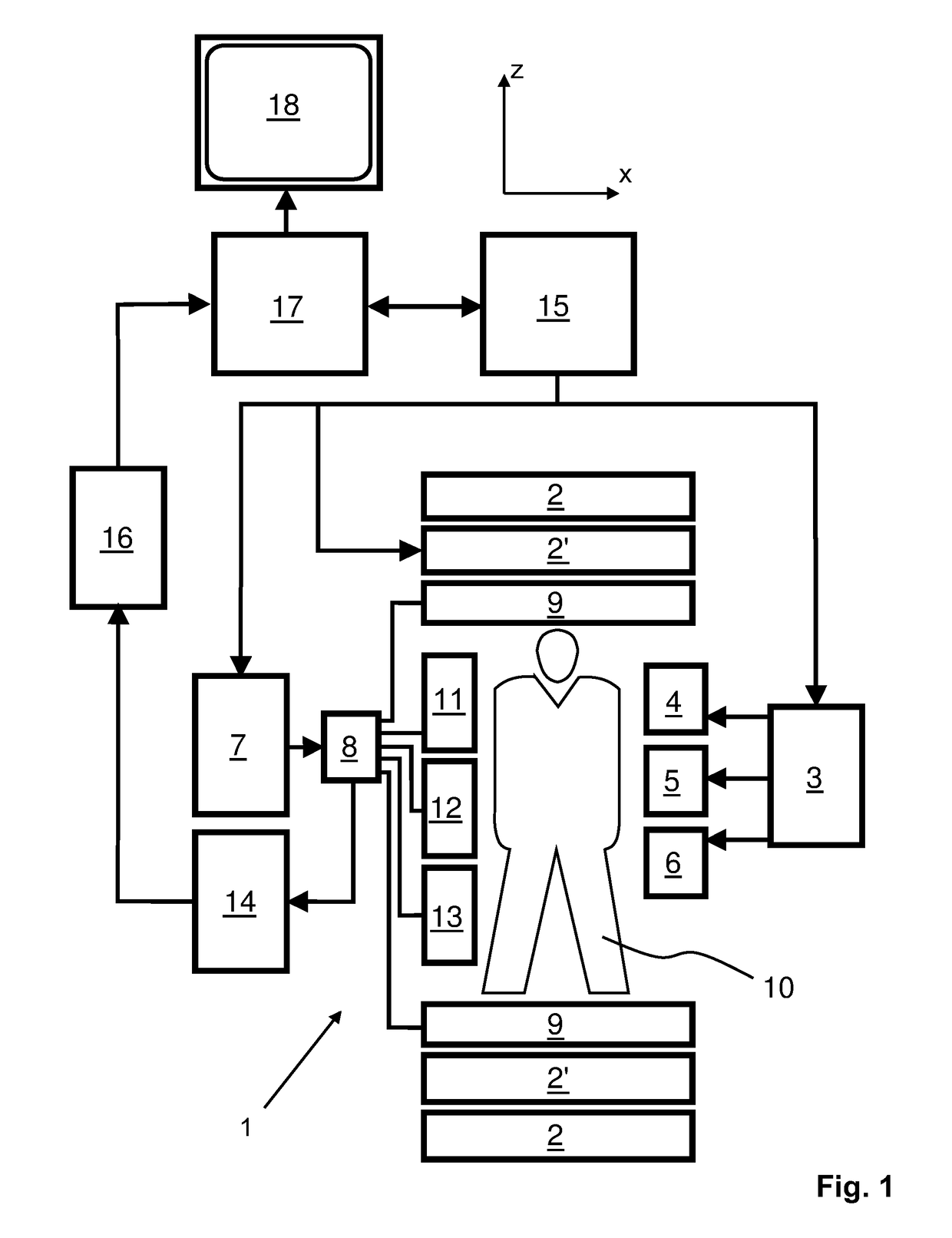
[0039]With reference to FIG. 1, a MR device 1 is shown. The device comprises superconducting or resistive main magnet coils 2 such that a substantially uniform, temporally constant main magnetic field B0 is created along a z-axis through an examination volume. The device further comprises a set of (1st, 2nd, and—where applicable—3rd order) shimming coils 2′, wherein the current flow through the individual shimming coils of the set 2′ is controllable for the purpose of minimizing B0 deviations within the examination volume.

[0040]A magnetic resonance generation and manipulation system applies a series of RF pulses and switched magnetic field gradients to invert or excite nuclear magnetic spins, induce magnetic resonance, refocus magnetic resonance, manipulate magnetic resonance, spatially and otherwise encode the magnetic resonance, saturate spins, and the like to perform MR imaging.

[0041]More specifically, a gradient pulse amplifier 3 applies current pulses to selected ones of whole-...


PUM


Abstract

The invention relates to a method of MR imaging near metal parts using SEMAC. It is an object of the invention to provide an improved MR imaging technique that is sufficiently fast and robust against susceptibility effects. The invention proposes to apply a weaker slice-selection magnetic field gradient (Gslice) for reduction of ripple-artefacts near metal parts or to apply undersampling in the slice-selection direction of the SEMAC sequence or to apply both these aspects. According to one aspect of the invention, a sparsity constraint is used to make the reconstruction of the undersampled MR images more stable. Moreover, the invention relates to a MR device (1) and to a computer program to be run on a MR device (1).

Description

CROSS-REFERENCE TO PRIOR APPLICATIONS[0001]This application is the U.S. National Phase application under 35 U.S.C. § 371 of International Application No. PCT / IB2014 / 058069, filed on Jan. 06, 2014, which claims the benefit of U.S. patent application No. 61 / 756,500, filed on Jan. 25, 2013. These applications are hereby incorporated by reference herein.FIELD OF THE INVENTION[0002]The invention relates to the field of magnetic resonance (MR) imaging. It concerns methods of MR imaging of at least a portion of a body. The invention also relates to a MR device and to a computer program to be run on a MR device.BACKGROUND OF THE INVENTION[0003]Image-forming MR methods which utilize the interaction between magnetic fields and nuclear spins in order to form two-dimensional or three-dimensional images are widely used nowadays, notably in the field of medical diagnostics, because for the imaging of soft tissue they are superior to other imaging methods in many respects, do not require ionizing ...

Claims


Application Information

Patent Timeline
Patent Type & Authority Patents(United States)
IPC IPC(8): G01R33/565G01R33/44G01R33/28G01R33/561
CPCG01R33/5659G01R33/288G01R33/445G01R33/56563G01R33/5615G01R33/56536G01R33/5611
Inventor JURRISSEN, MICHEL PAUL JURRIAANDEN HARDER, JOHAN MICHIELBLUME, ULRIKE ANDREADE WEERDT, ELWINVAN IJPEREN, GERRIT HENDRIK
Owner KONINKLJIJKE PHILIPS NV
Why Eureka
Try Eureka