Apparatus and method for intragastric balloon with in situ adjustment means

a technology of in situ adjustment and abdominal cavity, which is applied in the direction of diaphragms, obesity treatment, surgery, etc., can solve the problems of prolonging the procedure, difficult in situ balloon manipulation to visually locate the valve, and causing satiety for overweight patients, so as to improve the effect of weight loss over time, slow weight loss, and volum

Active Publication Date: 2008-07-17
APOLLO ENDOSURGERY INC
View PDF99 Cites 135 Cited by
  • Summary
  • Abstract
  • Description
  • Claims
  • Application Information

AI Technical Summary

Benefits of technology

[0021]Because of its in situ adjustability, the balloon can be filled over the duration of its placement, if desired, to improve weight loss over time. If weight loss is slow, the surgeon can add volume to the device to take up greater space in the stomach and encourage early satiety. With the improved weight loss over time, the device would benefit from a longer placement time while having a variable fill volume. To improve the duration of adjustability, the shell is preferably made from a highly acid resistant material such as diphenyl silicone, polytetrafluoroethylene (PTFE), modified PTFE, silicon-polyurethane elastomers, high density polyethylene (HDPE), low density polyethylene (LDPE) or other acid resistant shell materials. The shell may also be coated with a coating such as parylene to improve the acid resistance of device.

Problems solved by technology

The balloon occupies space in the stomach, thereby leaving less room available for food and creating a feeling of satiety for the overweight patient.
One difficulty frequently encountered with this type of intragastric balloon is locating the valve when the balloon is already in the stomach and the surgeon attempts to reinsert the filler tube for the purpose of adding or removing fluid from the balloon.
Those skilled in the art will readily appreciate that manipulating the balloon while in situ to visually locate the valve is rather difficult, and the process of searching for the valve undesirably prolongs the procedure.
Even with the incorporation of such tabs into current intragastric balloon designs, the surgeon may still encounter significant difficulty in finding the valve for filling or removing fluid from the balloon.
And even after the valve has been visually located, it is often still difficult or awkward for the surgeon to reinsert the filler tube into the example valve.
The balloon may be slippery and positionally unstable.
Another problem associated with the heretofore known methods and devices is that following placement of the intragastric balloon, a patient may experience nausea from the interaction of the recently placed gastric balloon within the stomach.

Method used


Image

  • Apparatus and method for intragastric balloon with in situ adjustment means
  • Apparatus and method for intragastric balloon with in situ adjustment means
  • Apparatus and method for intragastric balloon with in situ adjustment means

Examples


example 1

In Vivo Adjustment of a Balloon With a Button / Valve Assembly

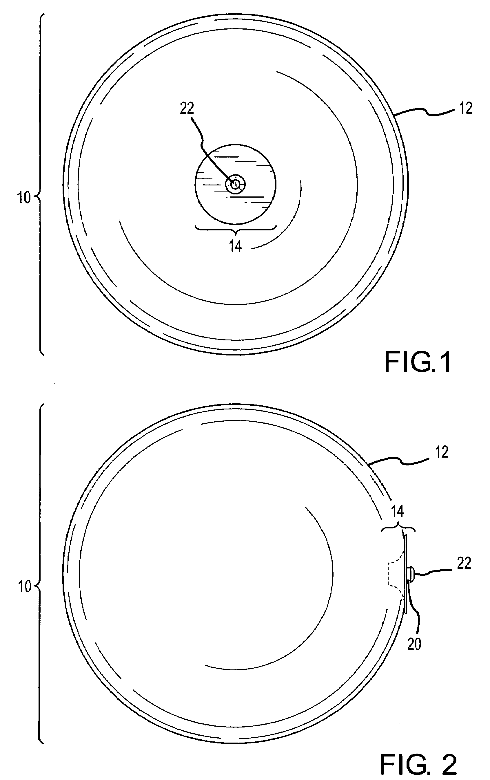
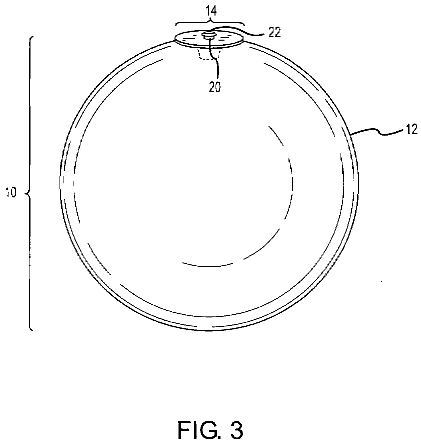
[0050]In this example, the surgeon performs an in vivo adjustment of an intragastric balloon that has been previously implanted in a patient. In this example, the surgeon wishes to add more fluid to a previously implanted intragastric balloon that includes a grasping button / valve assembly, such as that shown in FIGS. 4 & 5. The gastroscopic instrument of this example is equipped with a camera, a needle for adding fluid, and a grasping tool for capturing the button of the button / valve assembly located on the surface of the intragastric balloon.

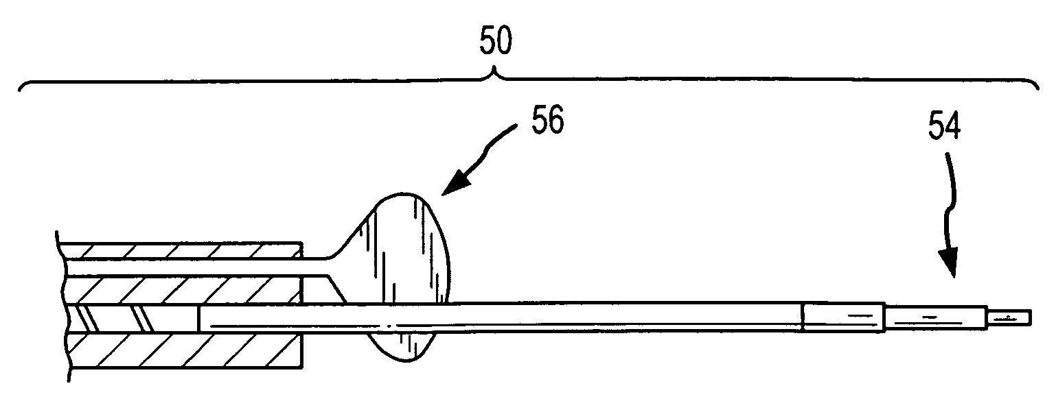
[0051]The patient is anesthetized, and the surgeon begins the procedure by inserting a gastroscopic instrument into the stomach. Using a camera located on a specially-configured gastroscopic instrument, such as that shown in FIGS. 6 & 7, the surgeon maneuvers the gastroscopic instrument into position to grasp the grasping button / valve assembly. The surgeon positions grasping tool 52 (F...

example 2

In Vivo Adjustment of a Balloon With Self-Sealing Shell

[0055]In this example, the surgeon performs an in vivo adjustment of an intragastric balloon that has been previously implanted in a patient. In this example, the surgeon wishes to add more fluid to a previously implanted intragastric balloon that includes a self-sealing portion, such as that shown in FIG. 9, wherein a grasping tab is located on the self-sealing portion. The gastroscopic instrument of this example is equipped with a camera, a needle for adding fluid, and a grasping tool for capturing the grasping tab located on the surface of the intragastric balloon.

[0056]The patient is anesthetized, and the surgeon begins the procedure by inserting a gastroscopic instrument into the stomach. Using the camera located on a specially-configured gastroscopic instrument, such as that shown in FIGS. 6 & 7, the surgeon maneuvers the gastroscopic instrument into position to grasp the grasping tab 24. The surgeon positions the grasping...

example 3

In Vivo Adjustment of a Balloon With Dual-Hemisphere Configuration

[0060]In this example, the surgeon performs an in vivo adjustment of an intragastric balloon that has been previously implanted in a patient. In this example, the surgeon wishes to add more fluid to a previously implanted intragastric balloon that is of a dual-hemisphere configuration, such as that shown in FIG. 11. The gastroscopic instrument of this example is equipped with a camera, a needle for adding fluid, and a grasping tool for capturing the intragastric balloon.

[0061]The patient is anesthetized and the surgeon begins the procedure by inserting a gastroscopic instrument into the stomach. Using the camera located on a specially-configured gastroscopic instrument, such as that shown in FIGS. 6 & 7, the surgeon maneuvers the gastroscopic instrument into position to fit over one of the hemispheres of the balloon. The surgeon positions the grasping tool of the gastroscopic instrument in its “open” position”. The ph...


PUM


Abstract

An intragastric balloon and method of adding and/or removing fluid therefrom are disclosed. The intragastric balloon includes a shell, a grasping tab, and a self-sealing portion. A gastroscopic instrument which includes a grasping tool is inserted into the stomach of a patient and used to grasp the grasping tab located on the surface of the shell. Using the grasping tool, the intragastric balloon is securely fastened to the gastroscopic instrument. A filling instrument, such as a needle, is advanced through the self-sealing portion of the shell for the filling or deflating of the intragastric balloon. After inflation or deflation is complete, the filling instrument is then withdrawn from the balloon and the gastroscopic instrument withdrawn from the stomach.

Description

BACKGROUND OF INVENTION[0001]1. Field of the Invention[0002]The present invention is directed to devices and methods that enable inflatable intragastric balloons used for the treatment of obesity to be filled, and in particular to devices and methods that enable the intragastric balloon to be filled, adjusted, or deflated while the device itself is in the stomach.[0003]2. Description of the Related Art[0004]Intragastric balloons are well known in the art as a means for weight loss and treating obesity. One such inflatable intragastric balloon is described in U.S. Pat. No. 5,084,061 and is commercially available as the BioEnterics Intragastric Balloon System (sold under the trademark BIB®). These devices are designed to provide therapy for moderately obese individuals who need to shed pounds in preparation for surgery, or as part of a dietary or behavioral modification program.[0005]The BIB® System, for example, consists of a silicone elastomer intragastric balloon that is inserted i...

Claims


Application Information

Patent Timeline
Patent Type & Authority Applications(United States)
IPC IPC(8): A61M29/00
CPCA61F5/0036A61F5/003
Inventor BIRK, JANEL
Owner APOLLO ENDOSURGERY INC
Why Eureka
Try Eureka