Ocular collar stent for treating narrowing of the irideocorneal angle

a technology of irideocorneal angle and ocular collar, which is applied in the field of human eye surgically implanted devices, can solve the problems of essentially ineffectiveness, reduced ability to actuate change and focus, and progressively flaccid focusing apparatus, so as to restore some level of accommodation and restore some phakic eye accommodative ability

Inactive Publication Date: 2019-04-23
RENKE PETER
View PDF55 Cites 0 Cited by
  • Summary
  • Abstract
  • Description
  • Claims
  • Application Information

AI Technical Summary

Benefits of technology

[0023]A stent device implanted into the anterior chamber angle could provide various structural elements and forces to restore some accommodative ability in the phakic eye. The stent must have precise anatomical placement to achieve this. The positioning of the stent is essentially along the internal foundation of the cornea. In this position it can induce forces radiating outward and anteriorly along the posterior cornea base. The forces would translate to a re-positioning of the accommodative apparatus to a more posterior placement. In this position the eye can restore some level of accommodation. The forces applied at the corneal base can also be adjusted to manipulate corneal curvature and change refractive error.

Problems solved by technology

The focusing apparatus becomes progressively more flaccid over time.
The musculature and feedback mechanism remain intact, but the ability to actuate change and focus diminishes throughout life, until it is essentially totally ineffective.
The youthful eye has excessive ability to accommodate in the order of tenfold of what is generally required.
This surplus erodes over time, and usually reaches the threshold of being initially problematic at 40 to 45 years old.
The degradation continues to progress and usually renders the eye with complete inability to sustain any near focus by the age of 60.
These predictable changes result in an anterior shifting of the lens and focusing apparatus.
The matrix of the crystalline lens becomes cloudy and impairs vision.
This typically becomes problematic enough to require cataract surgery between the ages of 70 to 80.
This increase in diameter results in outward displacement of the zonule attachments.
At the stage prior to surgery, the intact aged eye has to maintain function with very unfavourable displaced and compressed anatomy.
This shift results in restriction of flow based on the structural change.
The natural architecture of this porous tissue cannot perform its function well under compression.
Thus, a significant portion of the population does not live long enough to require cataract surgery based on vision needs.
Early POAG cases are likely, in part, the inability of the eye's drainage system to facilitate a safe pressure clearing time.
These therapies are hindered by costs, compliance and side effects.
Managing drug side effects in an elderly population is difficult.
Trabeculectomy is very invasive surgery with considerable side effects.
Scarring of the treated area is the greatest risk for failure.
Other risks include infection, haemorrhaging, cataract formation, and hypotony.
With hypotony, if the pressure remains too low for prolonged periods maculopathy and possible vision loss my result.
Both procedures have very limited success and usually still require chronic paramedical use All procedures which involve surgical trauma to compromised tissues must contend with the tissue's natural healing mechanism.
The early positive response often renders a compromised eye at greater risk for failure.
It is also difficult to be confident that the stent has penetrated the tissues as required to be effective.
All glaucoma treatments and variations of treatments have notable disadvantages and limited success.
Medication has significant side effects and surgery results in tissue trauma.
In both cases, the cost of treatment is high.
Procedural short term benefits can often result in additional costs associated with failure and complications.
Trauma and scarring will inevitably compound the regenerative process.
This results in corneal edema leading to decreased vision and potentially vision loss.
Both are effective procedures, but often have complications and limited duration of effectiveness.
In these cases, the stimulation can be beneficial or detrimental if over stimulated.
Such excessive power to focus would translate into significant forces induced by the ciliary muscles and accommodative apparatus.
Flashes or sparks of light in the absence of true light are indicative of tension at retinal adhesions and present with greater risk.
The feedback mechanisms to accommodate also remains, but the cortical stimulation to accommodate is met with a lack of response that ultimately can produce an over active muscular response of the accommodative apparatus.
The action and reaction do not balance and cause instability.
The retinal adhesion will be put under significant traction resulting in the inevitable PVD and risk of retinal detachment.
The relationship is complex and it is difficult to isolate the components of the accommodative mechanism.
This procedure is still utilized, but in limited circumstances due to the larger more invasive incision needed.
These are not truly reversible techniques because tissue is removed with the laser.
Intacs have not gained significant market share despite being marketed as a reversible procedure.
However, the eye is dynamic and changes in refractive status do have predictable patterns as well changes that cannot be anticipated.
At this stage there is no effective accommodation.
These lenses will continue to develop, but presently have limited success.

Method used


Image

  • Ocular collar stent for treating narrowing of the irideocorneal angle
  • Ocular collar stent for treating narrowing of the irideocorneal angle
  • Ocular collar stent for treating narrowing of the irideocorneal angle

Examples


Embodiment Construction

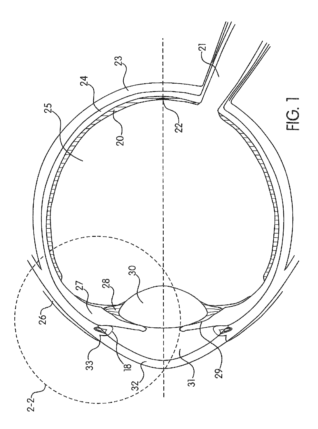
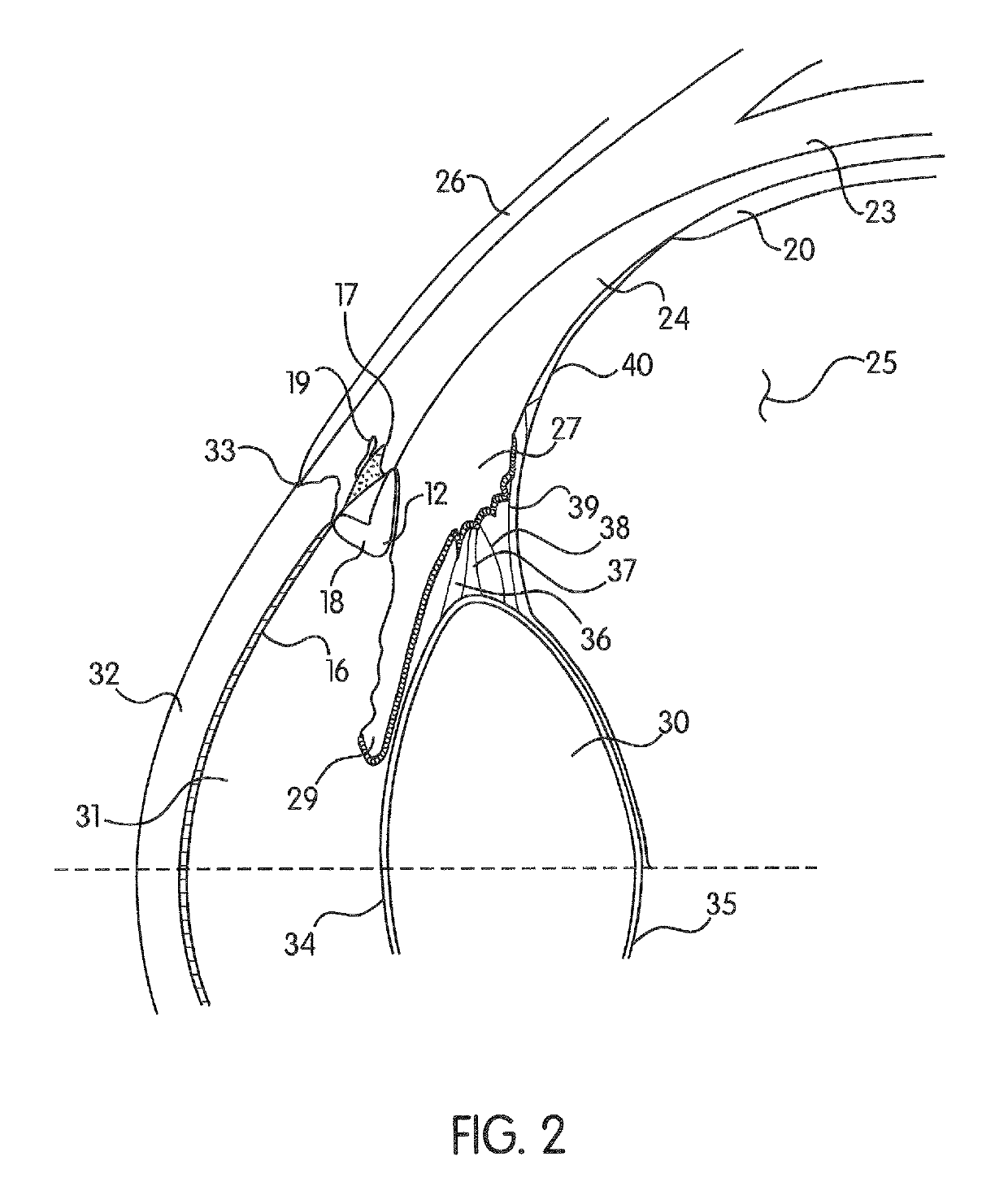
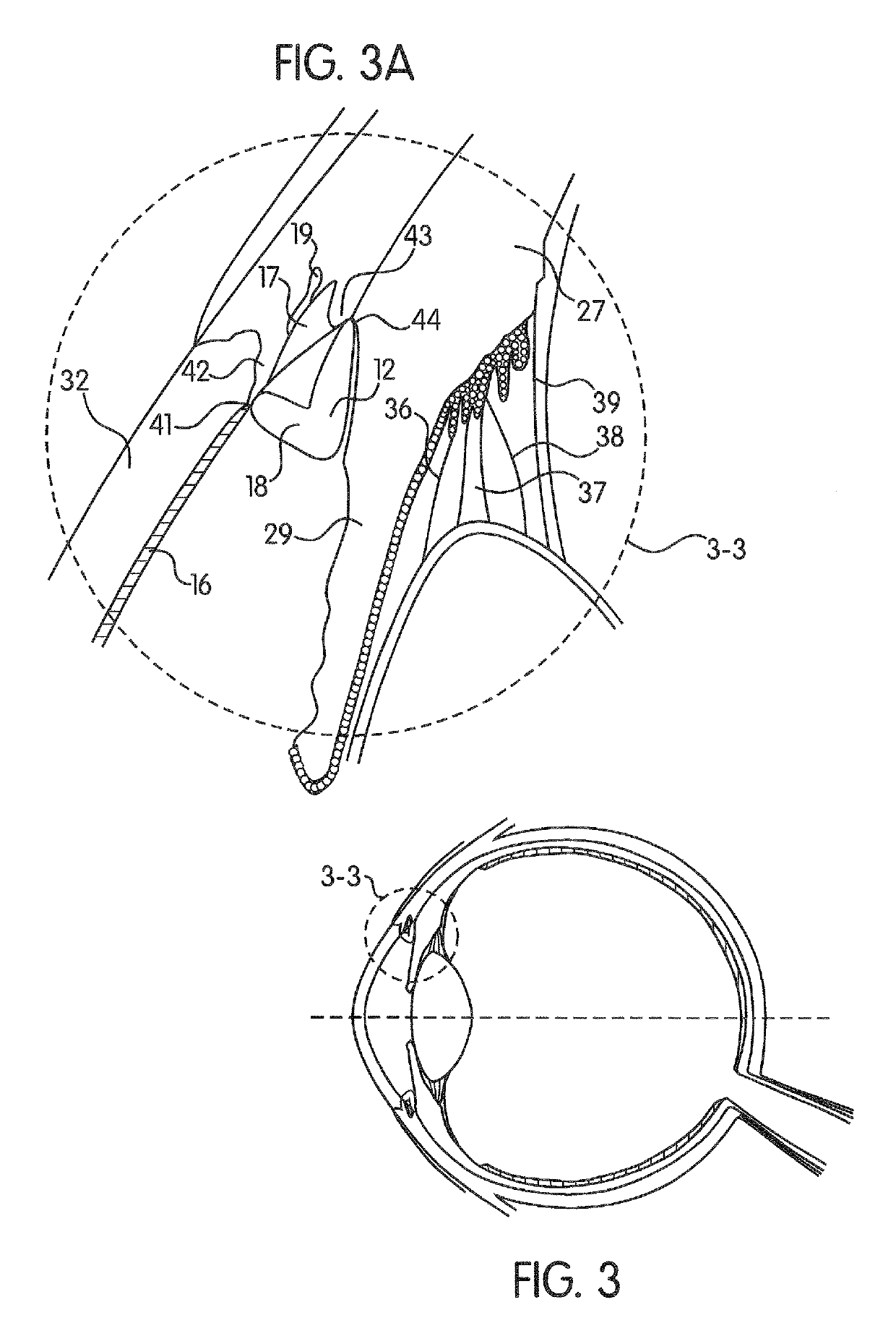
[0096]The illustrations generally show preferred embodiments of the devices used to treat predictable structural ocular aging changes of the anterior segment of the human eye. While the descriptions present various embodiments of the device(s), it should not be interpreted in any way as limiting the invention. Furthermore, modifications, concepts and applications of the inventions embodiments are to be interpreted by those skilled in the art as being encompassed, but not limited to the illustrations and descriptions herein.

[0097]The following description is provided to enable those skilled in the art to make and use the described embodiments contemplated for carrying out the invention. Various modifications, equivalents, variations, and alternatives, however, will remain readily apparent to those skilled in the art. Any and all such modifications, variations, equivalents, and alternatives are intended to fall within the spirit and scope of the present invention. Further, for purpose...


PUM


Abstract

An ocular stent for insertion in an anterior chamber of an eye is provided. The stent facilitates the restoration of the structure of an irideocorneal angle of the anterior chamber for treating structural changes from ocular aging. The stent includes: a continuous annular central portion; an anterior portion protruding in an anterior direction from the central portion and being configured to provide variable contact around a circumference of the stent with a surface of a transition zone between a trabecular meshwork and a corneal endothelium of the eye; and a posterior portion extending in a posterior direction from the central portion, the posterior portion being configured to contact a portion of a peripheral iris of the eye. A method for stabilizing the irideocorneal angle of the anterior chamber using a stent is also disclosed herein.

Description

CROSS REFERENCE TO RELATED APPLICATION[0001]This application is a continuation-in-part of U.S. patent application Ser. No. 13 / 960,416, filed Aug. 6, 2013, which claims priority to U.S. Provisional Patent Application No. 61 / 680,453 filed on Aug. 7, 2012, each of which is hereby incorporated by reference in its entirety.BACKGROUND OF THE INVENTION[0002]Field of the Invention[0003]This invention relates broadly to a structure for insertion in the anterior mammalian eye for treatment related to natural, predictable aging changes. More particularly the invention relates to a surgically implanted device for the human eye. The device provides structural support to specific ocular anatomy as a means to counteract changes naturally brought about by aging.[0004]Description of the Related Art[0005]All human eyes go through physical changes as a result of the noiinal aging processes. These changes affect various structures of the eye. The human eye has three chambers. The largest is the vitreou...

Claims


Application Information

Patent Timeline
Patent Type & Authority Patents(United States)
IPC IPC(8): A61F2/00A61F2/14A61F9/007A61F2/82
CPCA61F2/15A61F9/00781A61F2/82
Inventor RENKE, PETER
Owner RENKE PETER
Why Eureka
Try Eureka