Method for preparing mammalian tissue for storage, implant, and transplant

a technology of collagen tissue and mammalian tissue, which is applied in the field of processing isolated mammalian collagen tissue, can solve the problems of unsuitable transplantation, unsuitable corneal tissue, and unsanitary conditions, and achieve the effect of reducing storage temperature and increasing supply of transplantable tissu

Active Publication Date: 2019-05-28
FORSELL JAMES H +2
View PDF5 Cites 2 Cited by
  • Summary
  • Abstract
  • Description
  • Claims
  • Application Information

AI Technical Summary

Benefits of technology

[0095]As noted above, one advantage of the present invention is that tissue may now be split and partial corneas can be stored to increase the supply of transplantable tissue from eye banks. Although it has always been possible to split or cut a cornea into any shape or size, it was never logistically feasible, and therefore not commercially viable or surgeon friendly. The limiting factors have been storage time, clarity, pliability, and / or consistency of the cornea itself. The present invention combines long term preservation of a clear corneal stroma stored at room temperature, thus allowing for multiple grafts of the same cornea to become commercially viable without being limited by the factors of storage time, sterility, clarity, or consistency. Such practicality and commercial viability of cutting a cornea into 4 or more pieces has not previously been achieved.
[0096]Types of grafts achievable by the method of the present invention comprise the following non-limiting examples (see FIGS. 9A-9G):
[0097]1. 1 graft—an intact full thickness stromal graft 94. In another exemplary embodiment, the cornea may be pre-cut with center aperture 901, e.g., to enable use with a keratoprosthesis implant. In another exemplary embodiment, a cornea may be a full thickness cornea (clear center) with a scleral rim (white portion of the eye) 902 for surgeons who prefer to cut their own graft (this provides a flexibility on diameter for tectonic (structural) and surface grafting.
[0098]2. 2 half grafts—cutting a cornea in half resulting in two half-moon-shaped full thickness grafts 92 (e.g., glaucoma tube valve application).
[0099]3. 2 split grafts—making a transactional cut through a cornea resulting in two split thickness grafts of equal shape 93 or otherwise desired 95, 96 length or shape (e.g., corneal overlay).
[0100]4. 4 grafts—combining the 2 previous cuts (2, 3) resulting in four half-moon-shaped, split thickness grafts (not shown in figures, but a combination of 92 and 93) (e.g., glaucoma tube valve application).

Problems solved by technology

With the passage of time, the corneal tissue becomes edematous, loses its transparency and, ultimately, becomes unsuitable for transplantation.
Depending on the method applied, these methods may lack adequate levels of sterility, compromise the corneal stromal matrix, compromise the corneal clarity, and compromise the corneal elasticity or some combination thereof.
There are numerous problems associated with the Visiongraft process.
The ramification of this is that there exists a lot to lot variability that may affect performance.
The gamma source, however, generates heat in the environment that will increase the temperature within the exposure chamber.
Typically, the gamma source will be exposed to the irradiation chamber (raised out of its water environment) most of the working day, which causes a buildup of heat.
This use of 3 dimensional arrangements is not optimal for corneal tissue as it is difficult to obtain a uniform dose throughout the container.
Gamma radiation also poses a potential risk to those workers who operate the gamma radiation facility.
Overall, gamma radiation is expensive due to the necessary shielding for employees because of the deeply penetrating gamma rays, long exposure time due to the dose rate, inability to turn off the fuel rods, as well as the cost of final storage and disposition of spent fuel rods.
The cost of the extra pdHSA required to fill such a prior art vial is also a concern.
In addition, glass vials, after exposure to gamma radiation, will turn brown.
This browning makes it even more difficult to view the transparent tissue inside the storage container, which will create problems in ensuring adequate quality control during tissue release and tissue location / removal at surgery.
Additionally, glass vials can break or leak during transport or in hospital surgical suites during an operation, causing potential harm to both those handling the tissue as well as the tissue itself.
Breakage or leakage can also lead to a delay of the surgical procedure as well as upcharges to the patient.

Method used


Image

  • Method for preparing mammalian tissue for storage, implant, and transplant
  • Method for preparing mammalian tissue for storage, implant, and transplant
  • Method for preparing mammalian tissue for storage, implant, and transplant

Examples


example 1

ical Study

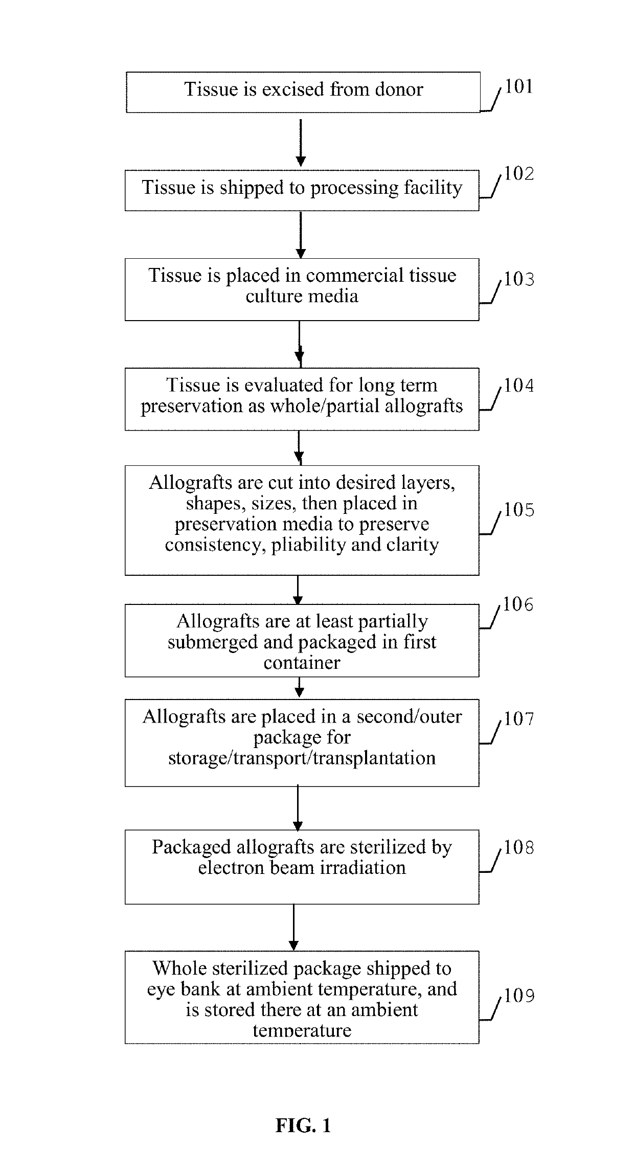
[0123]The MatTek EpiOcular™, a cultured, 3-dimensional in vitro human cell-based corneal model, was used to evaluate the potential of test articles to cause toxicity and / or ocular irritation of the new storage media after electron beam irradiation. The objective of this study was to assess the ocular irritation potential of the novel tissue storage media recombinant human albumin and compare it to 1). a known irritant, benzalkonium chloride and 2). the comparable product being offered for allograft transplants stored in pooled / processed human albumin. A positive control, 1% benzalkonium chloride (BC), was effective, reducing cell viability over three time periods. Therefore the positive control, 1% BC, is categorized as a severe irritant. Exposure of ocular tissues to rHSA had no effect on tissue viability up to and including the 300 min exposure. In addition, rHSA exposure did not induce a substantial release of IL-1α above what was observed with the negative control exce...

example 2

d Tissue Clarity

[0124]A pre and post-treatment comparison of 18 donor corneas was performed. The clarity of each tissue was evaluated and compared using dark field microscopy. Each cornea served as its own control, first as fresh tissue and then again following electron beam irradiation. Overall, there was a net change in clarity of −2.7% following treatment, p=0.002. The maintenance of corneal clarity utilizing this new processing and sterilization regimen is sufficient for clinical use.

example 3

d Tissue Histology

[0125]Corneal tissues subjected to the sterilization process were fixed in formalin and evaluated by an ocular pathologist after hematoxylin and eosin staining. The ocular pathological examinations were masked to fact that the tissue was subjected to a novel sterilization process. Tissue was deemed normal by histological examination. There were no identified differences between electron beam irradiated tissue and normal cornea.


PUM

PropertyMeasurementUnit
total volumeaaaaaaaaaa
temperatureaaaaaaaaaa
total volumeaaaaaaaaaa

Abstract

The present disclosure relates to methods and systems for processing isolated mammalian tissue for use in surgical grafting, implantation, or transplantation procedures. The method employs electron beam sterilization, the collagen tissue is partially submerged in rHSA (Recombinant Human Serum Albumin), the tissue is stored in specially designed packaging, and the tissue is stored at ambient temperatures without requiring refrigeration. The terminal sterilization using electronic beams takes place in the special packaging. This inventive technology offers advantages including being biologically safe, maintaining tissue integrity while minimizing size, increasing scalability, increasing shelf life, increasing the amount of tissue usable for transplantation, and reducing of costs associated with processing and storing tissue for long periods of time.

Description

CROSS-REFERENCE TO RELATED APPLICATIONS[0001]The present application is a Continuation-In-Part of U.S. patent application Ser. No. 14 / 314,084, filed Jun. 25, 2014, which claims priority to and incorporates fully by reference U.S. Provisional Patent Application Ser. No. 61 / 839,041, filed Jun. 25, 2013. Both related disclosures are incorporated herein by reference in their entireties.FIELD OF THE INVENTION[0002]The present invention relates to a method for processing isolated mammalian collagen tissue, and particularly mammalian corneal tissue, for use in surgical grafting, implantation, or transplantation procedures.BACKGROUND OF THE INVENTION[0003]Doctors have long been using organ and tissue transplantation to treat certain medial conditions. One example of tissue transplants is corneal tissue allografts. Corneal tissue allografts have been successfully conducted since 1905. Today, corneal tissue transplantation is used to treat poor vision due to corneal perforation, infection, sc...

Claims


Application Information

Patent Timeline
Patent Type & Authority Patents(United States)
IPC IPC(8): B65B63/08A61L2/00A61L27/36B65B55/16
CPCB65B63/08A61L2/007A61L27/3633B65B55/16A61L27/3691A61L2430/16A61L2202/23B65B5/045A61L2202/181
Inventor FORSELL, JAMES H.COLE, GERALD JOHNHOLIMAN, JEFFREY DOW
Owner FORSELL JAMES H
Why Eureka
Try Eureka