Diagnostic test for early stage cancer
a cancer diagnosis and early stage technology, applied in the field of cancer diagnostics, can solve the problems of incidentalomas, major problems threatening the quality and cost-containment of healthcare delivery, and invasive procedures, and achieve the effect of assessing the efficacy of cancer therapy
- Summary
- Abstract
- Description
- Claims
- Application Information
AI Technical Summary
Benefits of technology
Problems solved by technology
Method used
Image

Examples
example 1
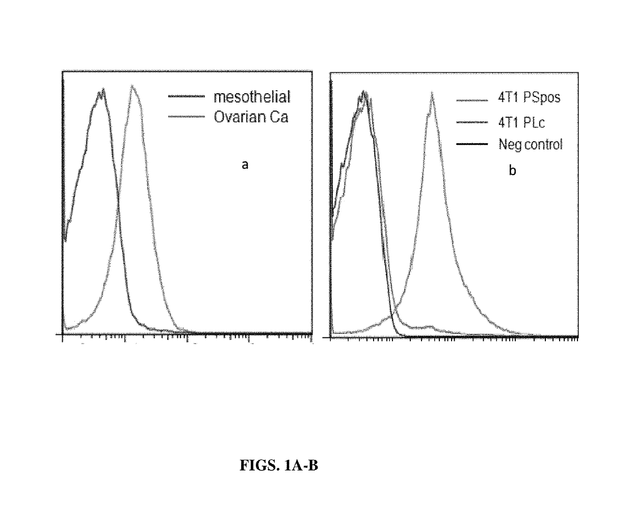
The Distribution of PS in the Membrane of Exsomes Derived from Normal Cells and from Tumor Cells
[0139]The present example, together with Table 2, show that in contrast to exosomes isolated from normal non-tumorigenic cells that do not express PS on the exosome surface membrane, exosomes isolated from tumor cells express relatively large measurable amounts of PS their membrane surface. These data suggest therefore that exosomes from normal or tumor cells can be distinguished from one another by tests that discriminate between the presence and absence of PS on the surface of exosomes.
[0140]A. Materials and Methods
[0141]Isolation of normal mesothelial cells and ovarian carcinoma cells from malignant ascites of an ovarian carcinoma patient. Ovarian tumor cell lines and mesothelial cell lines were established from 0.5 to 3.0 liters of ascites. The fluid was centrifuged and the resulting cell pellet was resuspended in medium and the tumor clusters were allowed to sediment while the mesoth...
example 2
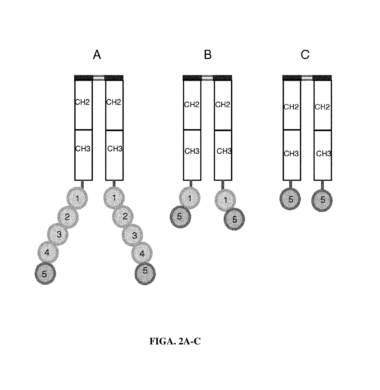
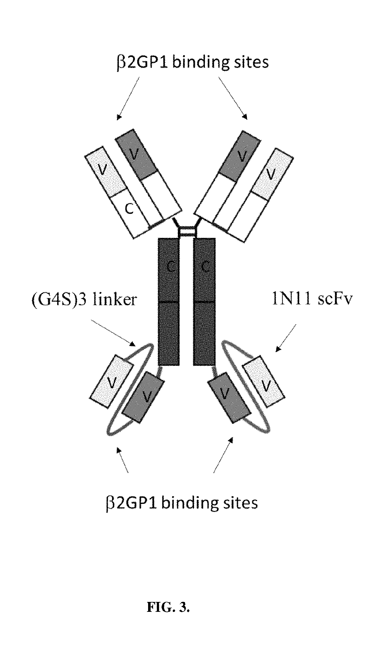
PS Binding Constructs
[0149]This example describes the design of various PS-binding constructs that selectively bind
[0150]PS-expressing tumor-derived exosomes. A series of PS binding proteins and constructs were tested as candidates for a high affinity capture moiety for use in the development of a sensitive and specific PS exosome assay. These included full-length β-2-glycoprotein 1 (β2GP1, also known as apolipoprotein H), a serum protein known to bind PS and various betabody constructs (FIGS. 2A-C).
[0151]Betabody constructs are dimeric (bivalent), PS binding proteins in which two β2GP1 polypeptides, each containing one or more domains of β2GP1, are attached to an antibody Fc region to create an antibody-like molecule. In a betabody, two β2GP1 polypeptides each containing at least the PS-binding domain 5 of β2GP1are attached to an antibody Fc region. Optionally, the two β2GP1 polypeptides in a betabody may additionally contain other β2GP1 domains, particularly domain 1, up to includ...
example 3
Assays Confirming PS Binding of PS Binding Proteins and PS Binding Constructs
[0154]The data presented in FIGS. 4-7 show the propensity of the various PS binding proteins to specifically bind PS that is incorporated into the membranes of artificially-generated vesicles containing PS. The results from all four assay systems (vesicle aggregation, ELISA, FACS and array analysis) confirm that the indicated PS-binding proteins / constructs can be used to quantify PS-expressing tumor exosomes present in patient blood.
[0155]Aggregation Assay. Multilamellar vesicles containing 50 mol % PS in PC were prepared in normal saline. These vesicles (50 μg) were mixed with 5 μg of the indicated binding agents and immediately placed on glass slides and photographed at 100× magnification. The extent of aggregation is an indication of the relative binding affinity of the various proteins / constructs. FIG. 4 shows weakest binding with KLSN, an N-terminal domain 5 containing betabody, intermediate binding wi...
PUM
| Property | Measurement | Unit |
|---|---|---|
| diameter | aaaaa | aaaaa |
| diameter | aaaaa | aaaaa |
| diameter | aaaaa | aaaaa |
Abstract
Description
Claims
Application Information
 - R&D
- Intellectual Property
- Life Sciences
- Materials
- Tech Scout
- Unparalleled Data Quality
- Higher Quality Content
- 60% Fewer Hallucinations
Browse by: Latest US Patents, China's latest patents, Technical Efficacy Thesaurus, Application Domain, Technology Topic, Popular Technical Reports.
© 2025 PatSnap. All rights reserved.Legal|Privacy policy|Modern Slavery Act Transparency Statement|Sitemap|About US| Contact US: help@patsnap.com