

Imaging apparatus for brachytherapy or biopsy
a technology for brachytherapy and biopsy, applied in the direction of therapy, catheter, ultrasonic/sonic/infrasonic image/data processing, etc., can solve the problems of inaccuracy of imaging, long time, and inability to accurately insert the introduction element, so as to achieve the effect of faster and more accurate inserting of the introduction elemen
- Summary
- Abstract
- Description
- Claims
- Application Information
AI Technical Summary
Benefits of technology
Problems solved by technology
Method used
Image

Examples
Embodiment Construction
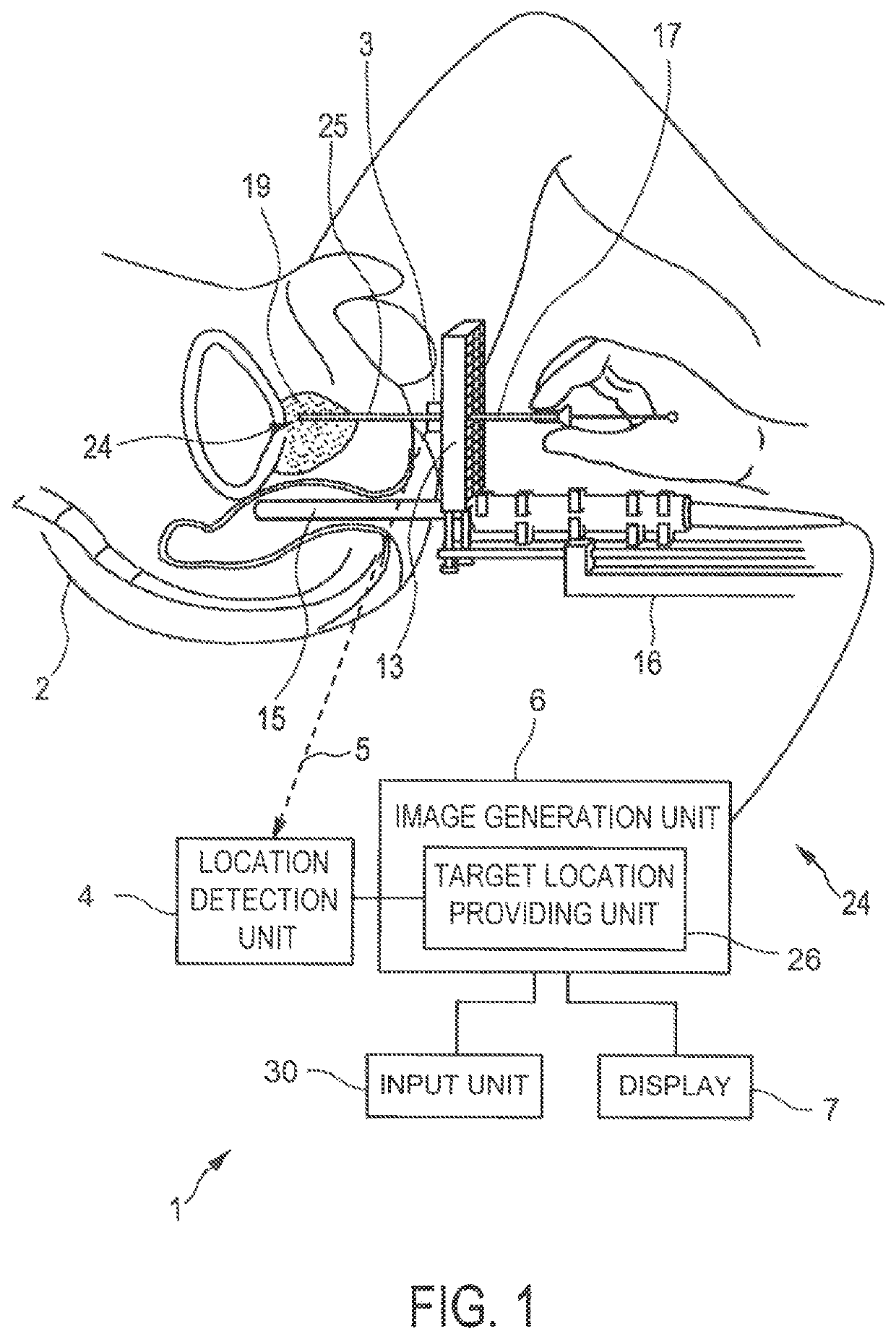
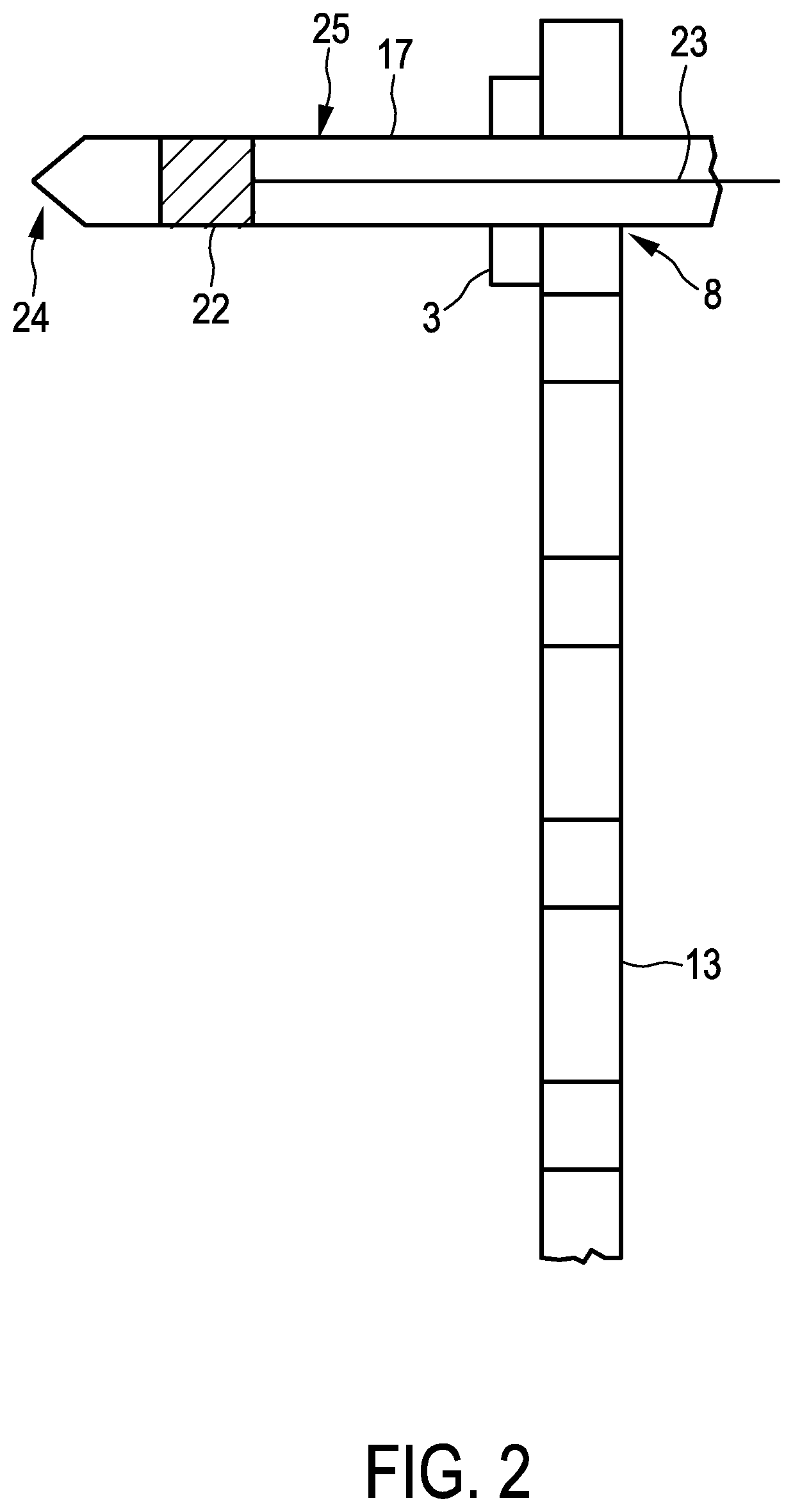
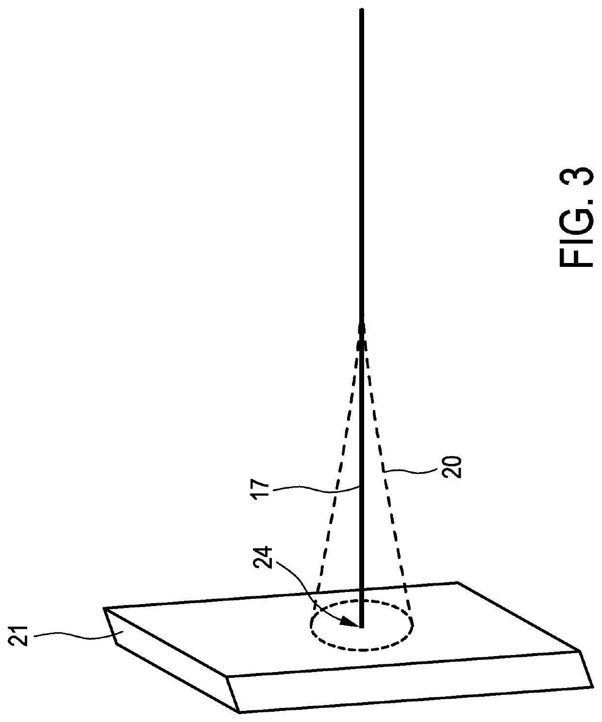
[0040]FIG. 1 shows schematically and exemplarily an embodiment of a brachytherapy system for performing a brachytherapy. The brachytherapy system 1 comprises an introduction element 17 for introducing a radiation source into a person 2. In this embodiment, the introduction element 17 is a needle, which is inserted into the prostate 19 of the person 2. During the insertion process an imaging apparatus 24 is used for imaging the introduction element 17 within the person 2.
[0041]The imaging apparatus 24 comprises a tracking unit 3, 4 for tracking the location of the introduction element 17 within the person 2, an imaging unit 6, 16 for generating an image showing an inner part of the person 2, which includes the tracked location of the introduction element 17, based on the tracked location, and a display 7 for displaying the image.
[0042]The introduction element 17 is manually inserted into the person 2, in particular, into the prostate 19, under image guidance. In particular, the imagi...
PUM
 Abstract
Description
Claims
Application Information