Determining image values in marked pixels of at least one projection image

a technology of projection image and image value, applied in image data processing, biological neural network models, computing models, etc., can solve the problems of further degradation of image quality, undermining the image quality of three-dimensional image dataset, etc., and achieve the effect of efficient and economical

Active Publication Date: 2021-03-02
SIEMENS HEALTHCARE GMBH
View PDF8 Cites 0 Cited by
  • Summary
  • Abstract
  • Description
  • Claims
  • Application Information

AI Technical Summary

Benefits of technology

[0014]Replacing defective image values or estimating and adding missing image values of marked pixels in projection images by inpainting methods affects a plurality of transformation values in the transformation images, while the use of a conventional inpainting method will, with high probability, produce no image values fulfilling the epipolar consistency conditions. However, by taking the epipolar consistency conditions into account in the determination of image values for the marked pixels in at least one projection image of the projection image set, the consistency is as far as possible maintained. For example, the combination of inpainting methods such as interpolation and / or artificial intelligence-based determination algorithms avoids inconsistent estimations and consequently, potential artifacts in the reconstructed three-dimensional image datasets.
[0015]In this case, the epipolar consistency conditions may generally be used in a simple and computationally efficient manner for the consistent estimation of image values that are to be determined. The image quality of the three-dimensional image dataset is increased, and artifacts may be avoided or reduced.
[0019]This enables epipolar consistency conditions to be selected that actually possess a relevance in terms of the image values to be determined in the marked pixels. In this way, epipolar consistency conditions that are less relevant or not relevant at all may be excluded from consideration, thus enabling a further estimation of the image values that are to be determined in an efficient and economical manner.
[0027]It may further be provided within the scope of the present embodiments that the determination algorithm performs an interpolating determination of the image values to be determined (e.g., in accordance with the normalized metal artifact reduction (NMAR) method). In this case, for example, a forward projection of an initially corrected volume may be called upon for an improved interpolation, in which case, however, a consistent estimation in accordance with the epipolar consistency conditions is not guaranteed. With this respect, the use of the epipolar consistency conditions as proposed here may also lead in combination with NMAR to substantially improved estimations for marked pixels.
[0029]In addition to the method, the present embodiments also relate to an X-ray device including a control device embodied to perform the method. All statements made in relation to the method according to the present embodiments may be applied analogously to the X-ray device, by which the already cited advantages may therefore likewise be obtained. For example, the X-ray device may be an X-ray device having a C-arm on which an X-ray source and an X-ray detector are arranged opposite each other. Alternatively, the X-ray device may be a computed tomography device in which, for example, at least one X-ray source and X-ray detector pair disposed within a gantry may be moved around the patient. The control device of such an X-ray device may be configured to estimate image values of marked pixels in the manner according to one or more of the present embodiments, which provides useful, high-quality functions for increasing the image quality of acquired image data and, for example, of three-dimensional image datasets already at the X-ray device itself. The control device may include at least one processor and / or at least one storage device and / or include a determination unit in order to determine image values to be determined using epipolar consistency conditions.

Problems solved by technology

The presence of metals in the examination region may seriously undermine the image quality of the three-dimensional image dataset.
However, an interpolation of the type lacks precision (e.g., in the case of truncated volumes; when the projection images are acquired by an X-ray device having a C-arm), thus leading to a further degradation in image quality.
However, the normalized metal artifact reduction requires a laterally untruncated scan of the object for the normalized interpolation act, since in this case, the values to be interpolated are estimated from numerically projected beams through the examination region.

Method used


Image

  • Determining image values in marked pixels of at least one projection image
  • Determining image values in marked pixels of at least one projection image
  • Determining image values in marked pixels of at least one projection image

Examples


Embodiment Construction

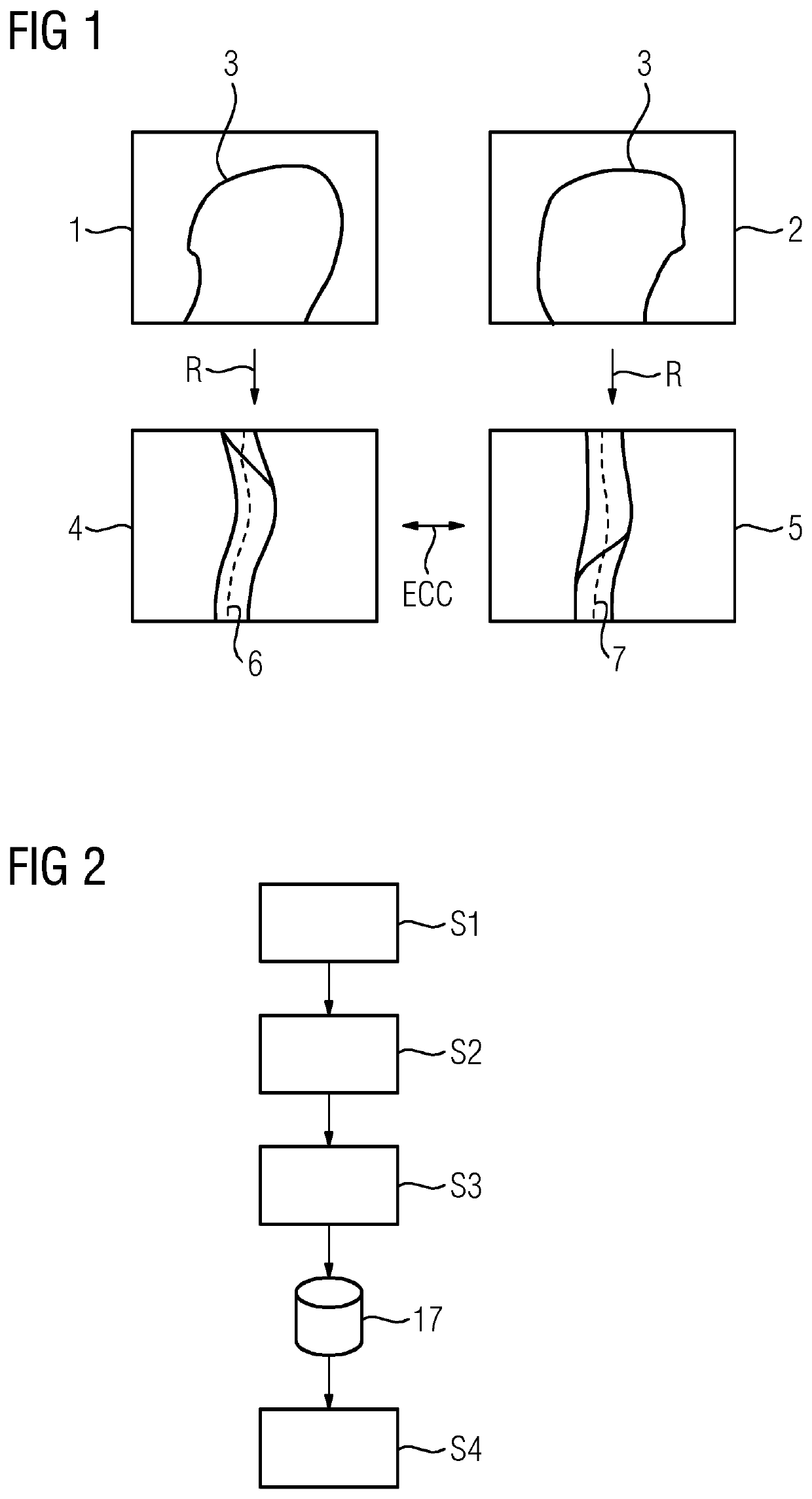
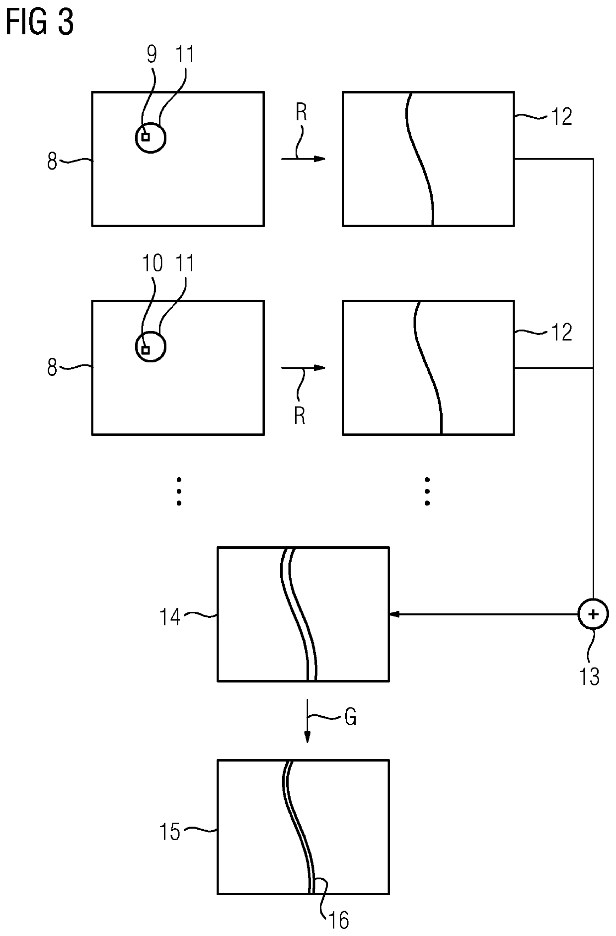
[0036]Exemplary embodiments of the method are presented below in relation to medical imaging and, more specifically, to metal artifact reduction in three-dimensional X-ray image datasets that are to be reconstructed. In this regard, as is generally known, a plurality of projection images of an examination region of the patient are acquired by an X-ray device using different projection geometries, each of which is described by a projection matrix (e.g., along a circular scanning trajectory). The projection images form a projection image set. Using the projection images, by applying corresponding reconstruction methods (e.g., filtered backprojection), a three-dimensional image dataset may be reconstructed from the two-dimensional projection images. This may, however, lead to artifacts if metal objects are present in the examination region. Metal objects, and consequently the cause of the metal artifacts, may therefore be computationally removed from the projection images by replacing ...


PUM


Abstract

A method for determining image values in marked pixels of at least one projection image is provided. The at least one projection image is part of a projection image set provided for reconstruction of a three-dimensional image dataset and acquired in each case using a projection geometry in an acquisition procedure. The image values are determined through evaluation of at least one epipolar consistency condition that is to be at least approximately fulfilled, that results from the projection geometries of the different projection images of the projection image set, and that requires the agreement of two transformation values in transformation images determined from different projection images by Radon transform and subsequent derivation as a condition transformation.

Description

[0001]This application claims the benefit of EP 18176803.7, filed on Jun. 8, 2018, which is hereby incorporated by reference in its entirety.BACKGROUND[0002]The present embodiments relate to determining image values in marked pixels of at least one projection image.[0003]It has become established practice in, for example, medical X-ray imaging to generate three-dimensional image datasets of an examination region of a patient based on an initial acquisition of X-ray projection images (e.g., along a circular trajectory) using different projection geometries. These two-dimensional (2D) X-ray projection images may be drawn upon as a basis for the reconstruction of a three-dimensional image dataset of the examination region (e.g., using the filtered backprojection method and / or the iterative (algebraic) reconstruction method). For example, 200 to 500 projection images may be acquired in this way using different projection geometries (e.g., projection angles) in the course of a circular p...

Claims


Application Information

Patent Timeline
Patent Type & Authority Patents(United States)
IPC IPC(8): A61B6/00G06N20/00G06N3/08G06T3/40G06T11/00
CPCA61B6/5205A61B6/4441G06N3/08G06N20/00G06T3/4007G06T11/006G06T11/008G06T11/005G06T2211/448
Inventor MANHART, MICHAEL
Owner SIEMENS HEALTHCARE GMBH
Why Eureka
Try Eureka