Imaging neurological disease

a neurodegenerative disease and imaging technology, applied in the field of radiopharmaceutical imaging of the brain, can solve the problems of compromising the utility and applicability of ‘normal’ average values reported, unable to establish a meaningful normal database that is built on data from more than one camera type, and unable to apply ‘correction factors’ with considerable effort,

Active Publication Date: 2021-07-06
GE HEALTHCARE LTD
View PDF0 Cites 1 Cited by
  • Summary
  • Abstract
  • Description
  • Claims
  • Application Information

AI Technical Summary

Problems solved by technology

It has therefore so far proved impossible to establish a meaningful normal database that is built on data from more than one camera type, without considerable effort to apply ‘correction factors’.
In the Hermes BRASS product, correction factors are applied but this significantly compromises the utility and applicability of the ‘normal’ average values reported.
A problem with this method is that average striatum binding ratio (SBR) and related values vary significantly between camera types, and even between the same camera type at different clinical sites—even when the same radiopharmaceutical is used.
The consequence is that the ‘average’ value of SBR cannot easily be transferred from one site to another as a comparator for the subject.
Furthermore, the conventional SBR calculates two ratios, so the error bar is compounded by the calculation.
Furthermore, whereas the diagnostic ‘dynamic range’ of SBR is decreased in patients of advanced age due to the natural reduction of striatal uptake even in normal subjects, this limitation is not present when comparing left:right uptake.

Method used


Image

  • Imaging neurological disease
  • Imaging neurological disease
  • Imaging neurological disease

Examples


examples

[0095]Embodiments of the present invention is illustrated by the non-limiting Examples detailed below. Examples 1 and 2 are theoretical Examples, whereas Examples 2-5 are based on clinical data.

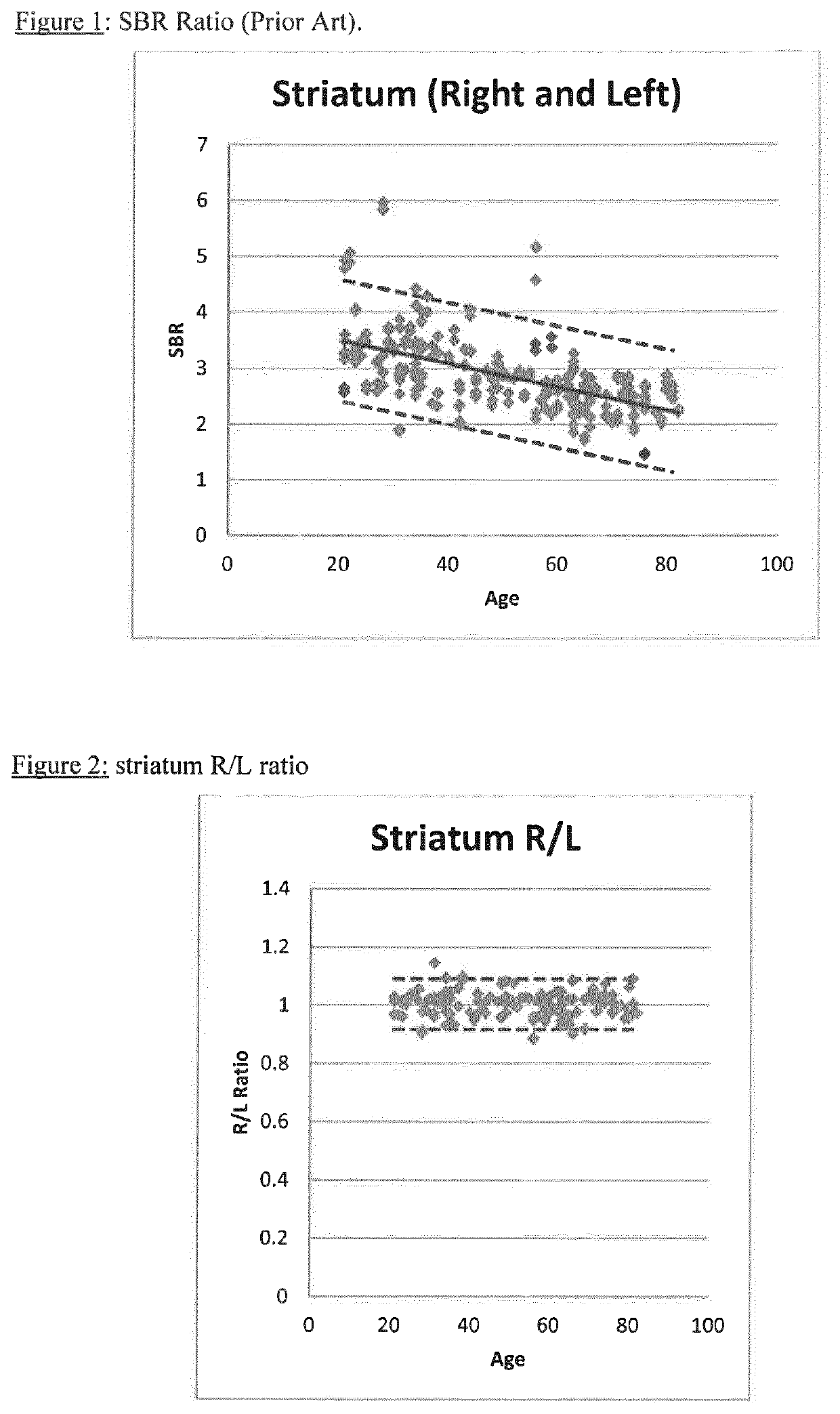
[0096]Example 1 is a comparative Example which shows the calculation of SBR using data from: (i) a range of camera manufacturers (Group A); (ii) a single manufacturer (GE; Group B) and (iii) a patient. The variation in Group A is so wide that there is almost no value in it for comparison with patient data. Even with Group B, the normal range is too wide to be useful for comparative purposes. This demonstrates the difficulties with the current methodology.

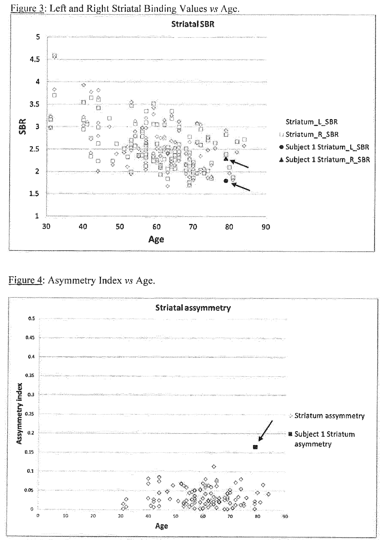
[0097]Example 2 applies the method of the present invention. In the normal population, for each individual subject the values for left and right are likely very close: a normal subject with striatum right=1.78 and striatum left=3.05 is inconceivable even though these represent −1 standard deviation and +1 standard deviation from the normal me...

example 1

Striatal Binding Ratios: Normal Range

Comparative Example

[0122]The European Association of Nuclear Medicine RESEARCH4LIFE© initiative (“EARL”) has been compiling a database of DaTSCAN images of normal human subjects since 2007 (see http: / / earl.eanm.org / cms / website.php?id= / en / projects / enc-dat.htm). The ENCDAT database includes 150 normal subjects—with data from a range of camera types. SBR was calculated for the 150 normal subjects (Group A):

[0123]SBR=(count⁢⁢density⁢⁢target⁢⁢ROI-count⁢⁢density⁢⁢reference⁢⁢ROI)count⁢⁢density⁢⁢reference⁢⁢ROI[0124]where:[0125]SBR=specific binding ratio or striatum binding ratio,[0126]ROI=region of interest.

Brain imaging data for n=37 normal subjects acquired on GE cameras in the ENCDAT database was used for the calculation of SBR (Group B). SBR for an illustrative patient from the ENCDAT database is also shown:

[0127]

TABLE 1SBR Striatum rightSBR Striatum leftNormal A (±1 SD)2.88 ± 0.652.88 ± 0.69Normal B (±1 SD)2.41 + / − 0.632.42 + / − 0.63

FIG. 1 shows the ...

example 2

Striatal Left / Right Binding Ratio

[0128]Using the method of the present invention, the data from Example 1 was used to calculate a striatum right:left ratio:

[0129]

TABLE 2StriatumSBR rightSBR leftRight:leftNormal2.88 + / − 0.652.88 + / − 0.691.00 + / − 0.04(+ / −1 SD)Normal range1.78-3.041.79-3.050.96-1.04(+ / −1 SD)

Table 3 below uses SBR data for normal subjects from the ENC DAT database. Three subjects from each of five different sites are shown, but the overall average is based on data from 11 sites:

[0130]

TABLE 3Stria-Puta-Anterior PosteriorSBRSBRtum Caudate menputamenPutamenStriatum Striatum R:LR:LR:LR:LR:LSiteRLratioratioratioratioratio14.934.791.031.011.041.011.113.233.221.001.030.990.961.073.153.091.021.051.001.000.9323.293.251.010.981.031.031.032.652.581.031.021.021.001.083.163.111.020.971.041.001.1832.742.601.051.051.051.001.182.692.940.920.890.930.911.001.911.881.021.031.000.981.0544.064.031.011.050.980.970.992.562.331.101.151.061.011.203.573.281.091.111.071.061.0752.702.621.031.071.0...


PUM

PropertyMeasurementUnit
2 dimensional imagingaaaaaaaaaa
volumeaaaaaaaaaa
tomographic imagingaaaaaaaaaa

Abstract

A method of imaging to permit calculation of left:right striatum uptake ratios is provided, and the degree of asymmetry used to assist in the diagnosis of neurological diseases. Also provided are a method of diagnosis, method of patient selection, and method of therapy monitoring using the imaging method, and software tools for use in the method.

Description

FIELD OF THE INVENTION[0001]Embodiments of the present invention relate to radiopharmaceutical imaging of the brain, in particular to dopamine transporter imaging of the striatum (or a portion thereof). A method of imaging to permit calculation of left:right striatum uptake ratios is provided, and the degree of asymmetry used to assist in the diagnosis of neurological diseases. Also provided are a method of diagnosis, method of patient selection, and method of therapy monitoring using the imaging method, and software tools for use in the method.BACKGROUND TO THE INVENTION[0002]Dopamine transporter imaging radiopharmaceuticals and brain imaging using such imaging agents have been described extensively in the art since the early 1990s. Laruelle et al [J. Cereb. Blood Flow Metab., 14, 982-984 (1994)] used the agent 123I-β-CIT and a ratio of striatal-to-background activity to give a figure proportional to dopamine transporter density.[0003]Tatsch et al [Quart. J. Nucl. Med., 56(1), 27-3...

Claims


Application Information

Patent Timeline
Patent Type & Authority Patents(United States)
IPC IPC(8): A61B6/00A61K51/04A61B6/02G16H50/20
CPCA61B6/481A61B6/02A61B6/501A61B6/5217A61K51/0455G16H50/20
Inventor ROSSER, MARKWOLBER, JAN
Owner GE HEALTHCARE LTD
Why Eureka
Try Eureka