
Method for detection and quantification of arterial calcification

a technology of arterial calcification and quantification method, which is applied in the field of method for detection and quantification of arterial calcification, can solve the problems of routine therapy, low detection rate, and high risk of disease and mortality for women, and achieve the effect of improving risk stratification

Active Publication Date: 2022-02-15
VOLPARA HEALTH TECH
View PDF9 Cites 0 Cited by
  • Summary
  • Abstract
  • Description
  • Claims
  • Application Information

AI Technical Summary

Benefits of technology

The present patent is about a method for improving the accuracy of breast density measurement by using BAC detection and quantification. This method creates a map of soft tissue that excludes calcified tissue, which is important for calculating breast density and assessing breast cancer risk. The method also takes into account important anthropomorphic features that can further improve risk stratification for various diseases. This is useful for breast cancer screening and prevention.

Problems solved by technology

There is known to be an association between arterial calcification and the risk of disease and mortality.
Although women are typically underserved in screening for cardiovascular-related disease.
A large proportion of cardiovascular events occur in women whose 10-year estimated risk of CVD is low, and thus guidelines would not routinely recommend therapy.
Consequently, there have been inconsistent findings.

Method used


Image

  • Method for detection and quantification of arterial calcification
  • Method for detection and quantification of arterial calcification
  • Method for detection and quantification of arterial calcification

Examples


Embodiment Construction

[0080]Herein are disclosed particulars of a method for the detection and quantification of breast arterial calcification, which uses information relating to tissue composition, breast anthropomorphic measures, and the application of these measurements as biomarkers for the prediction and stratification of risk of disease.

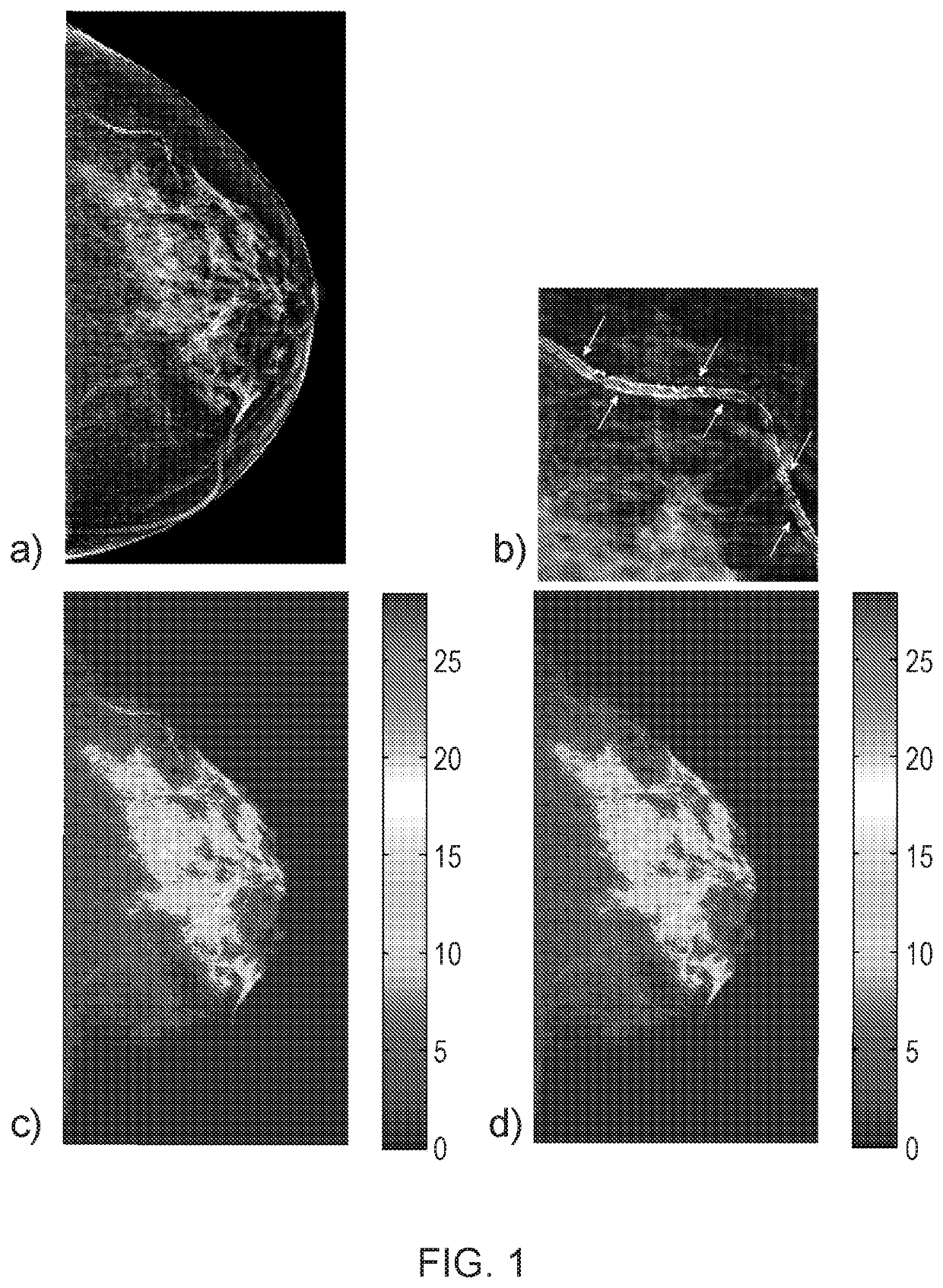
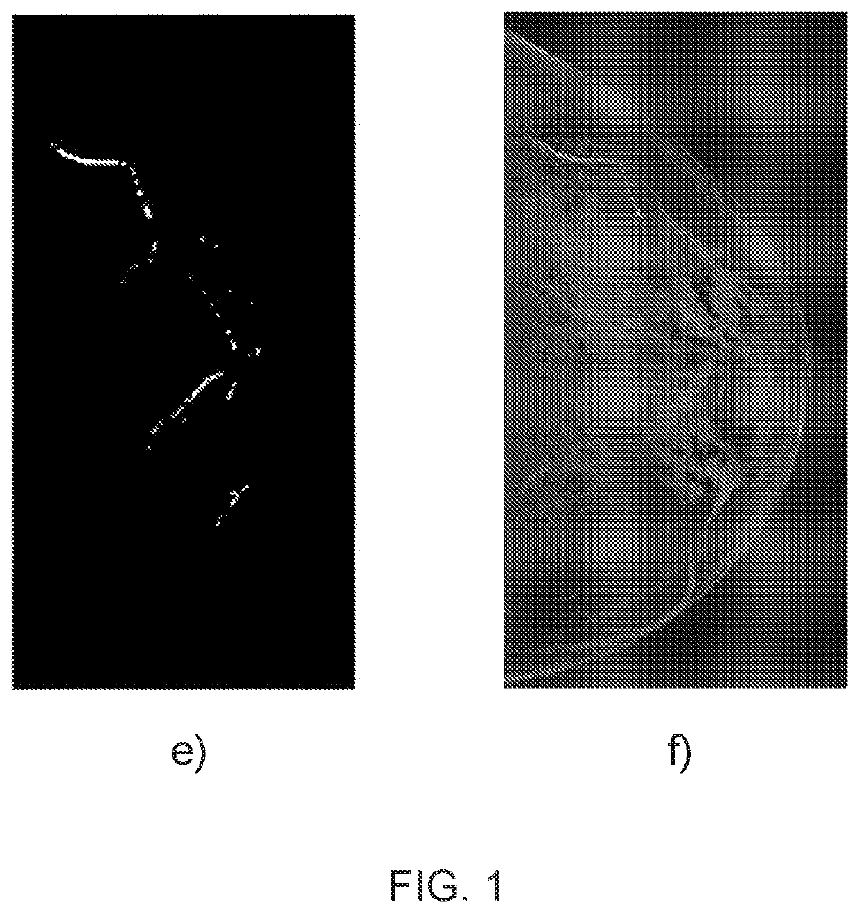
[0081]In an illustrative embodiment such as shown in FIG. 1, there is shown a mammogram which is a radiographic image of a breast. The radiographic image is transformed quantitatively to a tissue composition map.

[0082]The tissue composition map comprises a total amount of tissue in the breast associated with positions in the map. The tissue composition map may be illustrated by a height or a colour for each total amount of tissue for clear visualization. The positions and total amounts are also numerical quantities and may also be recorded or stored electronically for processing.

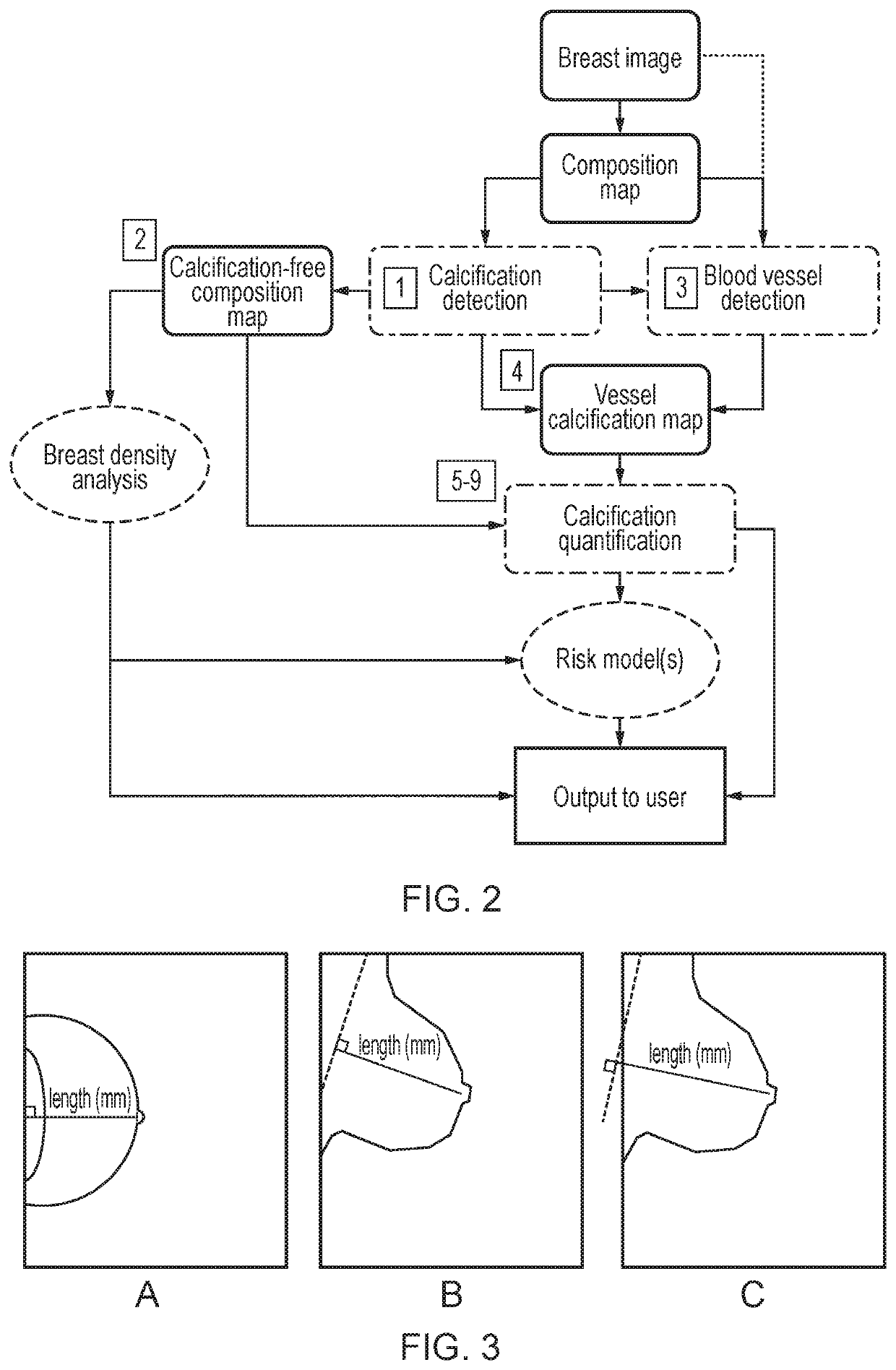
[0083]In an illustrative embodiment such as shown in FIG. 2, calcified arterial vessels ...


PUM


Abstract

Use of tissue composition and anthropomorphic measures in a method for the detection and quantification of arterial calcification in an organ is described for disease risk prediction and stratification. A radiographic image of an organ is transformed quantitatively to a tissue composition map indicating a total amount of organ tissue; a calcification map is generated indicating position in the tissue composition map of calcified tissue; calcification free tissue composition map is generated from the tissue composition map using the position of calcified tissue in the calcification map; a vessel map of the position of vessels in the tissue composition map is generated; and the vessel map is combined with the calcification map to generate a map of vessel calcification indicating the position of calcified vessels in the tissue composition map. Scores are based on arterial calcification in a breast which indicates disease in the breast and other organs.

Description

CROSS REFERENCE TO RELATED APPLICATIONS[0001]This application is a U.S. National Entry and claims priority to PCT International Patent Application No. PCT / IB2018 / 054462, filed Jun. 18, 2018, and entitled “METHOD FOR DETECTION AND QUANTIFICATION OF ARTERIAL CALCIFICATION”, which claims priority to New Zealand Patent Application No. 732921, filed Jun. 16, 2017, the entire disclosures of which are incorporated herein by this reference.FIELD OF THE INVENTION[0002]The present invention relates to use of tissue composition and anthropomorphic measures in a method for the detection and quantification of arterial calcification for disease risk prediction and stratification. More specifically the method relates to breast tissue composition and arterial calcification in the breast. The invention also relates to scores based on arterial calcification in a breast which indicates disease in the breast and other organs.BACKGROUND[0003]Vascular calcification is defined as “inappropriate and pathol...

Claims


Application Information

Patent Timeline
Patent Type & Authority Patents(United States)
IPC IPC(8): G06K9/00A61B6/00G06T7/12G16H50/30G06T7/00
CPCA61B6/502A61B6/5217G06T7/0012G06T7/12G16H50/30G06T2207/30068G06T2207/30101
Inventor HIGHNAM, RALPHHILL, MELISSA
Owner VOLPARA HEALTH TECH
Learn More