
Process for determining the infiltration of biological cells in a biological object of interest

a biological object and cell technology, applied in the field of biological image computer processing, to achieve the effect of robust curv

Active Publication Date: 2022-03-01
CENT NAT DE LA RECHERCHE SCI +5
View PDF3 Cites 0 Cited by
  • Summary
  • Abstract
  • Description
  • Claims
  • Application Information

AI Technical Summary

Benefits of technology

This process provides a robust and objective measure of immune cell infiltration, correcting for artifacts like holes and folds, and is applicable in both preclinical and clinical settings, producing consistent results regardless of tumor size or type, and is implementable automatically and quickly by a processing unit.

Problems solved by technology

One challenge is to obtain a repeatable and reproducible measure, producing objective results to assist the practitioner in making decisions, without replacing the practitioner.

Method used


Image

  • Process for determining the infiltration of biological cells in a biological object of interest
  • Process for determining the infiltration of biological cells in a biological object of interest
  • Process for determining the infiltration of biological cells in a biological object of interest

Examples


Embodiment Construction

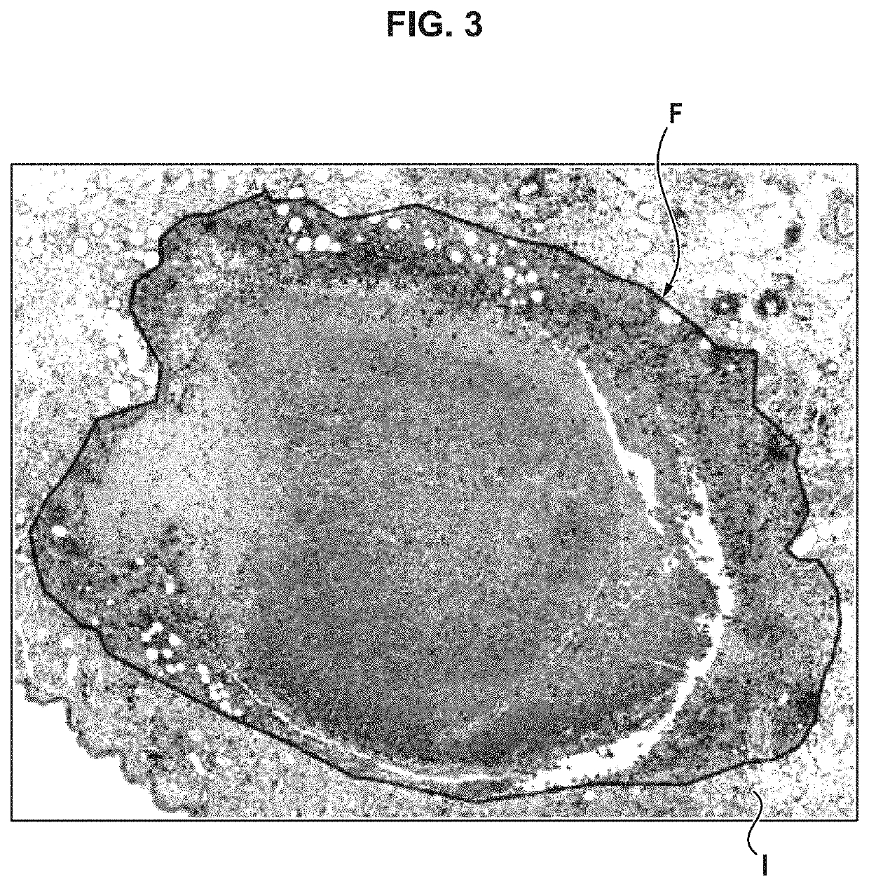
[0056]A process for determining the infiltration profile and an associated system are described below, for the particular case where the biological objects are tumors, for example cancerous tumors. The boundary of the biological object is therefore a tumor boundary in the following examples. It will be understood, however, that the invention can be used with the same advantages for the study of biological cell infiltration into biological objects other than tumors. For example, the following process can be applied to determine measures of infiltration of immune cells (such as lymphocytes) into a gland in the human or animal body, or into an organ transplanted into a patient, to determine whether there is rejection of the transplanted organ.

[0057]Hereinbelow, “histopathological image” means an image allowing a microscopic study of biological tissues, which can be used in histopathology in particular to monitor a disease. The biological tissues visible on such an image are most often ...


PUM


Abstract

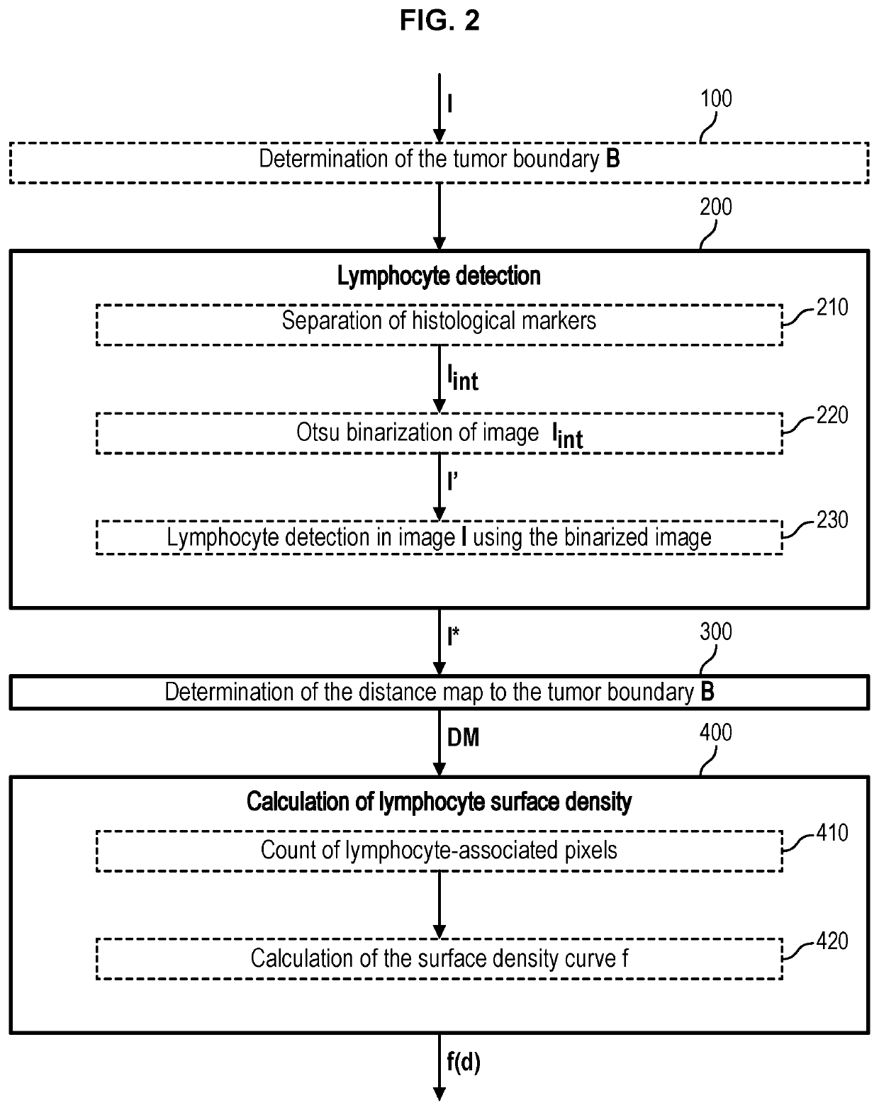
The invention relates to a process for determining an infiltration profile of biological cells of interest in a biological object of interest from a digital histopathological image of biological tissues, a histological stain having previously been applied to the biological tissues,comprising generating a biological cell detection image, pixels associated with the histological stain on the histopathological image being of a predetermined color on said image,determining a distance map comprising distance iso-curves to the boundary of the biological object,and, from the distance map, calculating a curve representative of the surface density of biological cells of interest as a function of distance to the boundary, by counting, for each boundary distance value, pixels that are both of the predetermined color on the detection image and located between the iso-curve associated with said distance value and the consecutive iso-curve.

Description

CROSS-REFERENCE TO RELATED APPLICATIONS[0001]The present application claims priority from French Application No. 1855513 filed Jun. 21, 2018, the disclosure of which is hereby incorporated herein by reference.TECHNICAL FIELD OF THE INVENTION AND STATE OF THE ART[0002]The invention belongs to the technical field of computer processing of biological images, preferably for applications in anatomic pathology.[0003]The malignancy of a tumor, for example cancer, can be diagnosed, and / or the efficacy of a treatment can be assessed, by analyzing microscopic images of a tumor sample. The sample collected is cut into sections, and these sections are stained to reveal certain types of biological cells. Microscopic images of the sections, including stained biological cells, can be used by an anatomic pathologist to establish the malignancy of the tumor, assess the patient's prognosis for survival, evaluate the efficacy of a treatment, etc.[0004]In particular, post-treatment immune cell infiltra...

Claims


Application Information

Patent Timeline
Patent Type & Authority Patents(United States)
IPC IPC(8): G06T7/90G06K9/00
CPCG06T7/90G06K9/00147G06T2207/10056G06T2207/30024G06T7/0012G06T2207/30096G06V20/698
Inventor LEMDANI, KATHIASEGUIN, JOHANNEDJIRO, ARMAND TIEDEEMILE, JEAN-FRANCOISKURTZ, CAMILLELOMENIE, NICOLAS
Owner CENT NAT DE LA RECHERCHE SCI
Learn More