
High resolution imaging apparatus and method

a high-resolution, imaging apparatus technology, applied in the direction of instruments, spectrometer detectors, particle separator tube details, etc., can solve the problems of unpredictability of sample breakdown and fragmentation, and achieve the effect of increasing speed

Active Publication Date: 2022-03-01
UNIVERSITY OF OTTAWA +1
View PDF6 Cites 2 Cited by
  • Summary
  • Abstract
  • Description
  • Claims
  • Application Information

AI Technical Summary

Benefits of technology

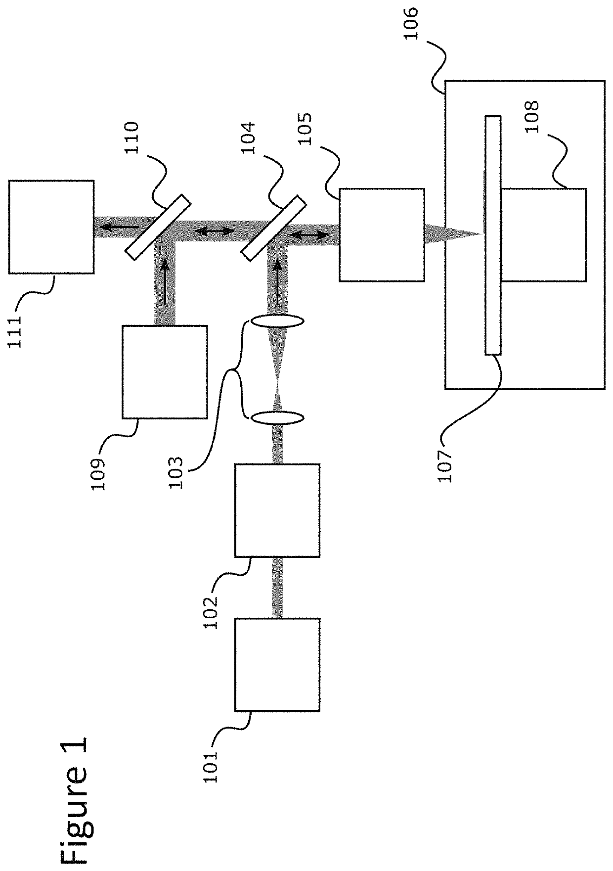
[0018]Although the detector system of the apparatus of the invention can detect many ions, most of these will be ions of the atoms that naturally make up the sample. In some cases, for example when analysing biological samples, the native element composition of the sample may not be suitably informative. This is because, typically, all proteins and nucleic acids are comprised of the same main constituent atoms, and so while it is possible to tell regions which contain protein / nucleic acid from those that do not contain such proteinaceous or nucleic acid material, it is not possible to differentiate a particular protein from all other proteins. However, by labelling the sample with atoms not present in the material being analysed under normal conditions, or at least not present in significant amounts (for example certain transition metal atoms, such as rare earth metals; see section on labelling below for further detail), specific characteristics of the sample can be determined. In common with IHC and FISH, the detectable labels can be attached to specific targets on or in the sample (such as fixed cells or a tissue sample on a slide), inter alia through the use of SBPs such as antibodies, nucleic acids or lectins etc. targeting molecules on or in the sample. In order to detect the ionised label, the detector system is used, as it would be to detect ions from atoms naturally present in the sample. By linking the detected signals to the known positions of the sampling of the sample which gave rise to those signals it is possible to generate an image of the atoms present at each position, both the native elemental composition and any labelling atoms. In aspects where native elemental composition of the sample is depleted prior to detection, the image may only be of labelling atoms. The technique allows the analysis of many labels in parallel (also termed multiplexing), which is a great advantage in the analysis of biological samples, now with increased speed due to the application of a laser scanning system in the apparatus and methods disclosed herein.

Problems solved by technology

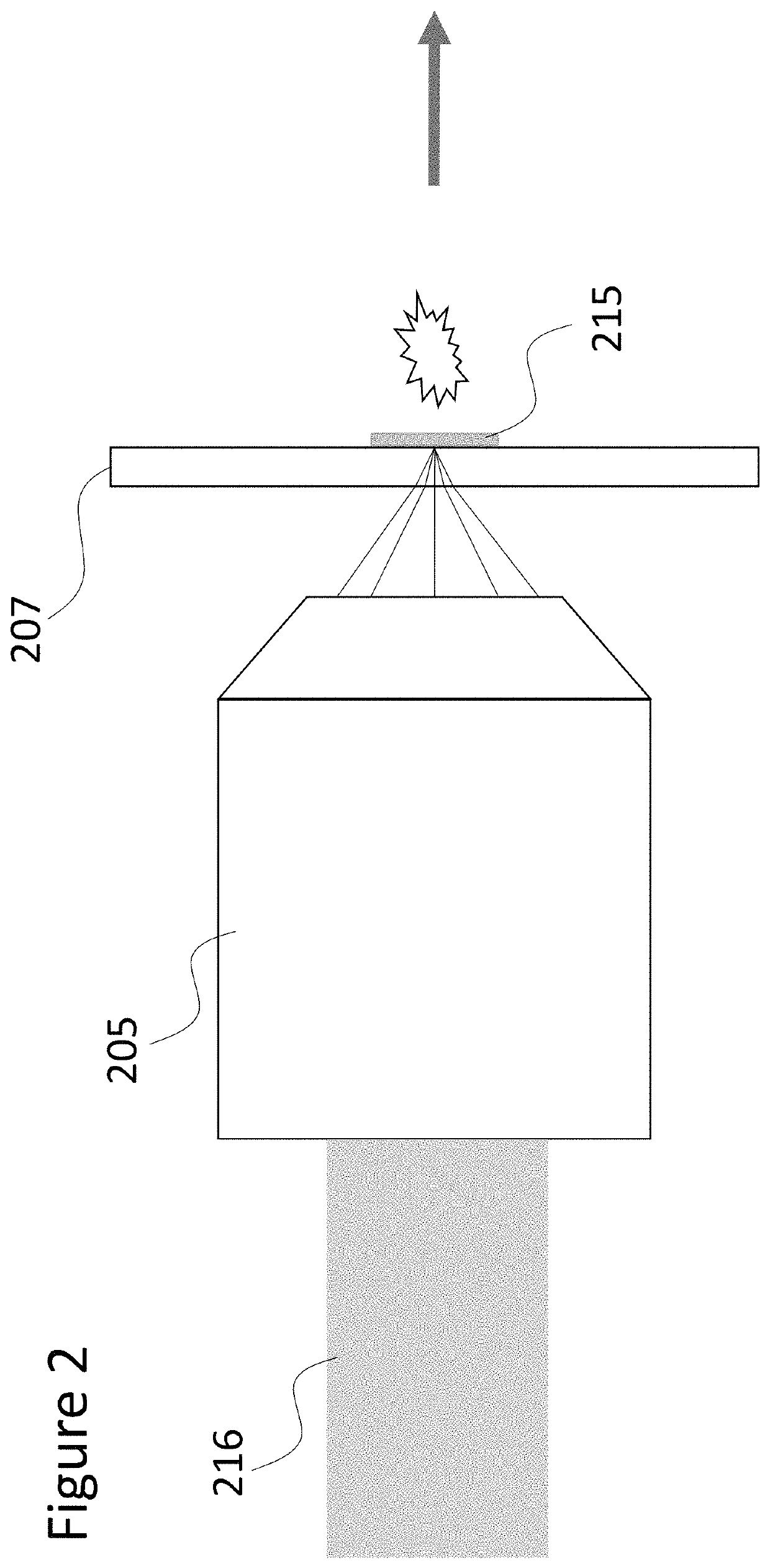
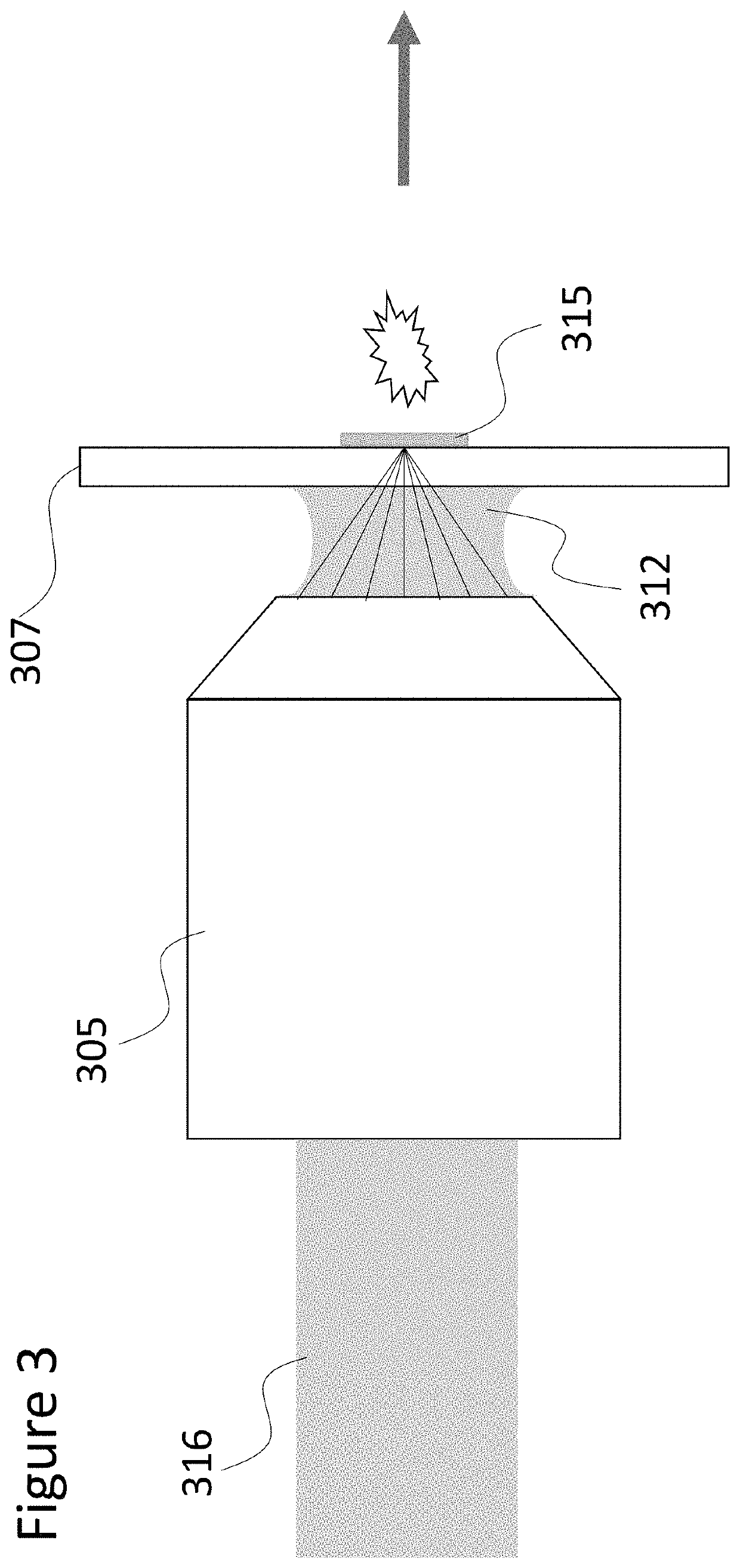
If the focal length is set such that the focus of the radiation is in the substrate rather than at its surface, this can lead to unpredictable breakdown and fragmentation of the sample.
This problem is caused in particular by the fact the inventors' finding that to use an immersion lens most effectively, the sample is best ablated in a manner that directs the laser radiation through the sample carrier.

Method used


Image

  • High resolution imaging apparatus and method
  • High resolution imaging apparatus and method
  • High resolution imaging apparatus and method

Examples


example 1

[0951]Protocol

[0952]The mouse gut was immersion fixed following organ harvesting using 2% PFA and 2.5% glutaraldehyde. The tissue was post-fixed with 2% Osmium Tetroxide for 1 hour. After dehydration the tissue sample was embedded in EPON resin and sectioned.

[0953]The 1000 nm tissue section was subjected to laser ablation and analyzed for the presence of osmium isotopes by Hyperion imaging system.

[0954]Results

[0955]FIG. 11 illustrates detection of abundant osmium isotopes (188Os, 189Os, 190Os and 192Os), as well as isotopes that are naturally low abundant (0.02% 184Os, 1.59% 186Os, 1.96% 187Os) present in the mouse gut tissue processed according to the standard immunoelectron microscopy protocols. The gut morphology is well defined with a dense outer layer of smooth muscle (upper left corner), mucosal epithelium with goblet cells and scattered stroma and immune cells beneath the epithelium.

[0956]Thus, electron microscopy 1 μm sections treated with 2% osmium tetroxide can be imaged f...


PUM

PropertyMeasurementUnit
wavelengthaaaaaaaaaa
spot sizeaaaaaaaaaa
thicknessaaaaaaaaaa

Abstract

The present invention relates to the high resolution imaging of samples using imaging mass spectrometry (IMS) and to the imaging of biological samples by imaging mass cytometry (IMCTM) in which labelling atoms are detected by IMS. LA-ICP-MS (a form of IMS in which the sample is ablated by a laser, the ablated material is then ionised in an inductively coupled plasma before the ions are detected by mass spectrometry) has been used for analysis of various substances, such as mineral analysis of geological samples, analysis of archaeological samples, and imaging of biological substances. However, traditional LA-ICP-MS systems and methods may not provide high resolution. Described herein are methods and systems for high resolution IMS and IMC.

Description

CROSS REFERENCE TO RELATED APPLICATIONS[0001]The present application claims the benefit of U.S. Provisional Application No. 62 / 686,341 filed Jun. 18, 2018 and entitled “HIGH RESOLUTION IMAGING APPARATUS AND METHOD,” and U.S. Provisional Application No. 62 / 792,759 filed Jan. 15, 2019 and entitled “HIGH RESOLUTION IMAGING APPARATUS AND METHOD,” both of which are hereby incorporated by reference in their entireties.FIELD OF THE INVENTION[0002]The present invention relates to the high resolution imaging of samples using imaging mass spectrometry (IMS) and the imaging of biological samples by imaging mass cytometry (IMC™).BACKGROUND[0003]LA_ICP-MS (a form of IMS in which the sample is ablated by a laser, the ablated material is then ionised in an inductively coupled plasma before the ions are detected by mass spectrometry) has been used for analysis of various substances, such as mineral analysis of geological samples, analysis of archaeological samples, and imaging of biological substan...

Claims


Application Information

Patent Timeline
Patent Type & Authority Patents(United States)
IPC IPC(8): H01J49/04G01N33/68G02B21/33H01J37/20H01J37/28H01J49/00H01J49/02H01J49/06H01J49/10H01J49/40H01J49/16H01J49/14
CPCH01J49/0463G01N33/6848G01N33/6851G02B21/33H01J37/20H01J37/28H01J49/0004H01J49/0031H01J49/025H01J49/0418H01J49/067H01J49/105H01J49/142H01J49/161H01J49/164H01J49/40G02B21/02G02B21/26G02B21/0032
Inventor CORKUM, PAULLOBODA, ALEXANDER V.
Owner UNIVERSITY OF OTTAWA
Learn More