Phosphoproteins in extracellular vesicles as candidate markers for breast cancer

a technology of phosphoproteins and vesicles, applied in the field of breast cancer candidate markers, can solve the problems of inability to keep the neural cytoskeletal structure organized, and no systematic method to decipher the correlation between phosphoproteins/glycoproteins and various cancer diseases

Active Publication Date: 2022-10-25
PURDUE RES FOUND INC
View PDF0 Cites 0 Cited by
  • Summary
  • Abstract
  • Description
  • Claims
  • Application Information

AI Technical Summary

Problems solved by technology

In Alzheimer's disease, due to misfoldings and abnormal conformational changes in tau protein structure, it is rendered ineffective at binding to microtubules and thus unable to keep the neural cytoskeletal structure organized during neural processes; in fact abnormal tau inhibits and disrupts microtubule organization and disengages normal tau from microtubules into cytosolic phase.
Despite the available knowledge to limited phosphoproteins and / or glycosignatures and diseases, to date, there is no systematic method to decipher the correlation between the phosphoproteins / glycoproteins and various cancer diseases.

Method used


Image

  • Phosphoproteins in extracellular vesicles as candidate markers for breast cancer
  • Phosphoproteins in extracellular vesicles as candidate markers for breast cancer
  • Phosphoproteins in extracellular vesicles as candidate markers for breast cancer

Examples


example 1

Identification of 9,643 Unique Phosphopeptides From Plasma Microvesicles and Exosomes

[0068]In this Example, we illustrated the preliminary screening of phosphopeptides from plasma microvesicles and exosomes.

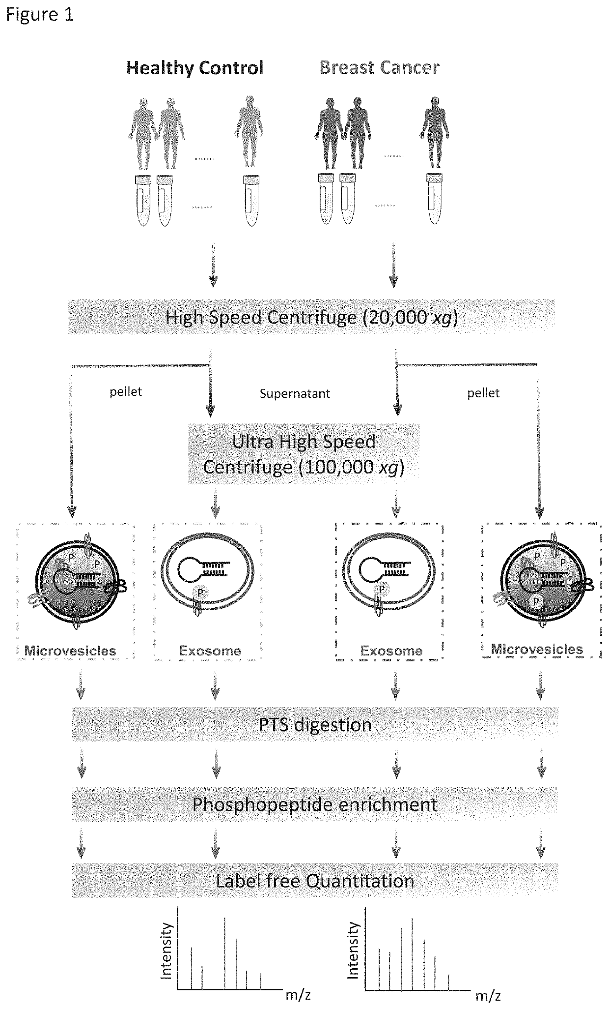
[0069]The workflow for the isolation of EVs, enrichment of phosphopeptides, and EV phosphoproteome analyses is illustrated in FIG. 1. Microvesicles and exosomes were isolated from human plasma samples through high-speed and ultra-high-speed centrifugations, respectively, an approach that has been employed in multiple studies and can effectively isolate EVs in good purity. For initial screening, the plasma samples were collected and pooled from healthy individuals (n=6) and from patients diagnosed with breast cancer (n=18). After lysis of EVs, proteins were extracted and peptides were generated using trypsin with the aid of phase-transfer surfactants (PTS) for better digestion efficiency and fewer missed tryptic sites. Phosphopeptides were enriched and followed by LC-MS / MS analyse...

example 2

Cancer Specific Phosphoproteins in EV

[0072]In this Example, we establish that looking the differences of EV phosphoprotein level from patient sample and healthy donor sample may identify a panel of phosphoproteins as the biomarker for diagnosing the disease at issue.

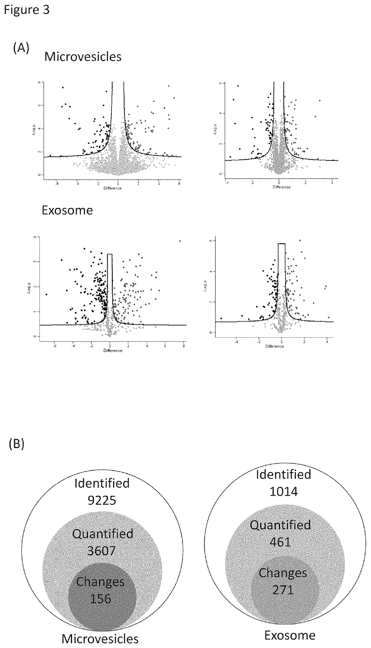
[0073]Label-free quantitation of phosphopeptides with probability score of phosphorylation site location over 0.75 was used to identify differential phosphorylation events in breast cancer patients from healthy individuals. We quantified 3,607 and 461 unique phosphopeptides and identified 156 and 271 phosphopeptides with significant changes (FDR<0.05 and S0=0.2) in microvesicles and exosomes, respectively (FIGS. 3A&B). Differential phosphorylation may be a result of changes in protein expression or changes in a particular site's phosphorylation. To distinguish these factors, we also performed the label-free quantitation of total proteomes for both microvesicles and exosomes (FIG. 3C). Altogether, we identified 1,996 prot...

example 3

Verification of Cancer Patient-Specific Phosphorylation Using PRM

[0076]In this Example we further validated the phosphoprotein biomarker candidate identified in Example 2.

[0077]Since breast cancer is extremely heterogeneous, the chance to identify a single diagnostic biomarker is likely rare. Instead, the identification of a panel of candidate markers that reflect the onset and progression of key disease-related signaling events would be feasible to offer better prognostic value. In effort to validate the differential phosphorylation of potential markers in cancer patients, we applied Parallel Reaction Monitoring (PRM) to quantify individual EV phosphopeptides in plasma from breast cancer patients and healthy individuals. Since phospho-specific antibodies suitable for construction of enzyme-linked immunosorbent assay (ELISA) are rarely available, targeted, quantitative MS approaches such as PRM and MRM (multi-reaction monitoring) are essential for initial validation. As a demonstrat...


PUM

PropertyMeasurementUnit
diameteraaaaaaaaaa
diameteraaaaaaaaaa
timeaaaaaaaaaa

Abstract

The state of protein phosphorylation and glycosylation can be key determinants of cellular physiology such as early stage cancer, but the development of phosphoproteins and / or glycoproteins in biofluids for disease diagnosis remains elusive. Here we demonstrate, for the first time, a strategy to isolate and identify phosphoproteins / glycoproteins in extracellular vesicles (EVs) from human plasma as potential markers to differentiate disease from healthy states. We identified close to 10,000 unique phosphopeptides in EVs by isolating from small volume of plasma samples. Using label-free quantitative phosphoproteomics, we identified 144 phosphoproteins in plasma EVs that are significantly higher in patients diagnosed with breast cancer than in healthy controls. Several novel biomarkers were validated in individual patients using Paralleled Reaction Monitoring for targeted quantitation. Similarly a group of glycoproteins in plasma EVs are identified. The study demonstrated that the development of phosphoproteins and / or glycoproteins in plasma EV as disease biomarkers is highly feasible and may transform cancer screening and monitoring.

Description

CROSS-REFERENCE[0001]This Application claims the benefit of U.S. Provisional Application 62 / 443,400, filed on Jan. 6, 2017. The disclosure of which is expressly incorporated herein entirely.STATEMENT OF GOVERNMENT SUPPORT[0002]This invention was made with government support under GM109626 and GM111788 awarded by the National Institutes of Health, and CHE1506752 awarded by the National Science Foundation. The government has certain rights in the invention.FIELD OF INVENTION[0003]This disclosure is related to a method to use small portions of patient body fluid sample to diagnose and monitor a pathological disease. Specifically the method uses non-invasive phosphoproteosome technology to identify a panel of phosphoproteins in extracellular vesicles as the biomarkers to diagnose and monitor breast cancer patient in a heterogeneous patient cohorts.BACKGROUND[0004]Extracellular vesicles (EVs) are produced by all domains of life including complex eukaryotes, both Gram-negative and Gram-po...

Claims


Application Information

Patent Timeline
Patent Type & Authority Patents(United States)
IPC IPC(8): G01N33/50G01N33/574G01N33/68
CPCG01N33/57415G01N33/57488G01N33/6842G01N33/6848
Inventor TAO, WEIGUO ANDYILYUK, ANTON B.ANDALUZ, HILLARYCHEN, I-HSUANPAN, LI
Owner PURDUE RES FOUND INC
Why Eureka
Try Eureka