
Visualization during closed-chest surgery

a closed-chest surgery and visualization technology, applied in the field of visualization during closed-chest surgery, can solve the problems of requiring one week of hospital recovery time, significant physical trauma to the patient, and weeks of convalescen

Inactive Publication Date: 2003-08-14
ATRICURE
View PDF17 Cites 46 Cited by
  • Summary
  • Abstract
  • Description
  • Claims
  • Application Information

AI Technical Summary

Benefits of technology

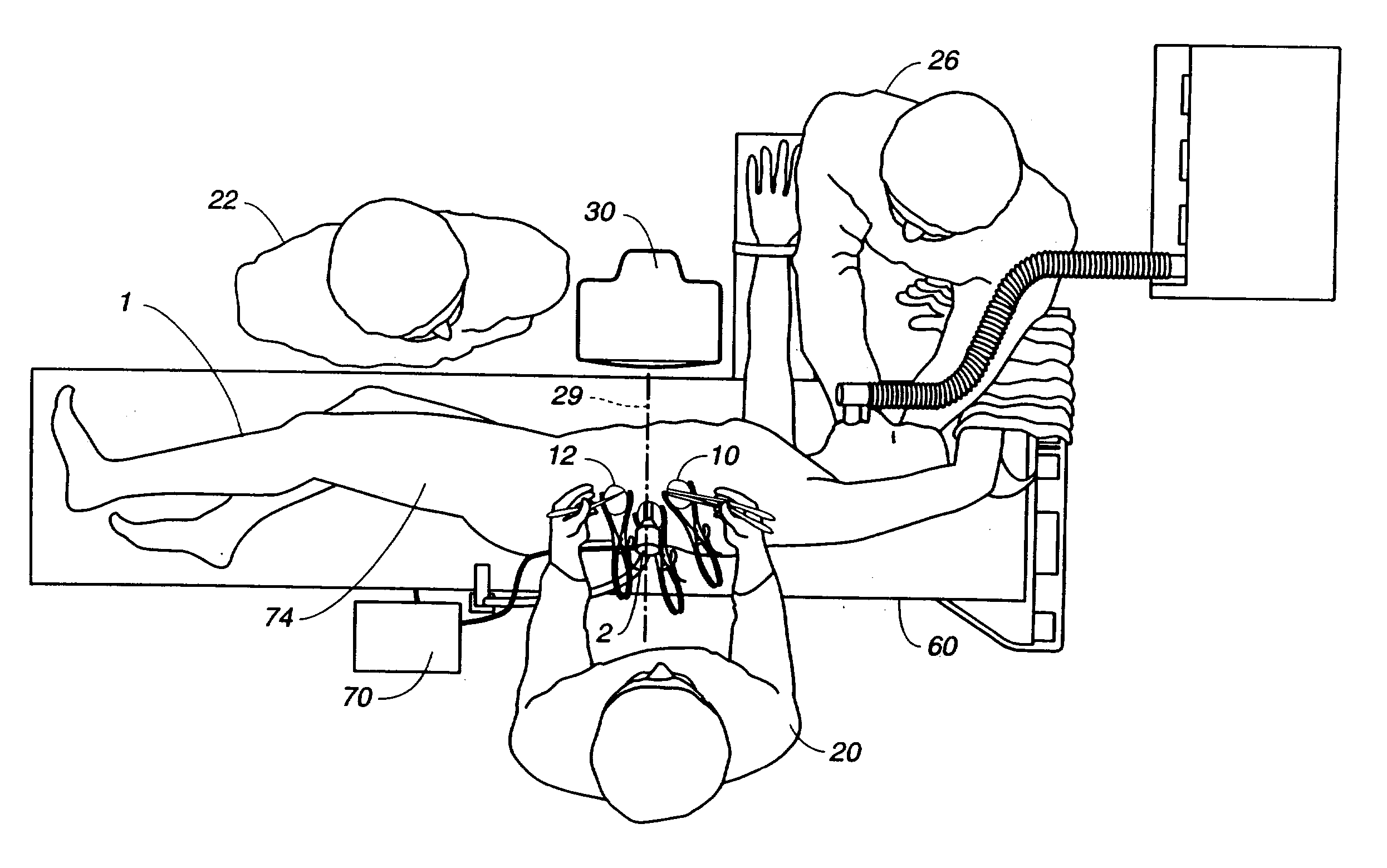
[0019] (b) positioning said draped monitor within the surgical field so that the surgeon can perform the internal surgical or diagnostic procedure and view it on said monitor so that the surgeon's visual access to the monitor correlates as closely as possible to the visual access of the input end of the viewing scope. Preferably the viewing monitor is a flat panel monitor that is on a movable cart having a sterilizable adjustable arm that allows the monitor to be positioned within the surgical field and adjusted to enhance the surgeon's visual alignment.
[0036] a view monitor capable of exhibiting the image gathered from the patient's cavity on a screen portion of the viewing monitor, wherein said camera is positioned at about a 90 degree angle to said nosepiece to minimize the distance said view scope extends out of said patient.
[0037] Still another aspect of this invention is a view scope having a light source, camera, and lens means, said view scope having the lens means positioned in a nosepiece designed to be inserted percutaneously into a patient's internal cavity to gather and transmit an image from the distal end of the lens means to said camera, wherein said camera is positioned at about a 90.degree. angle to said nosepiece to minimize the distance said view scope extends out of said patient.

Problems solved by technology

This technique causes significant physical trauma to the patient and can require one week of hospital recovery time and up to eight weeks of convalescence.
This can be very expensive in terms of hospital costs and disability, to say nothing of pain to the patient.
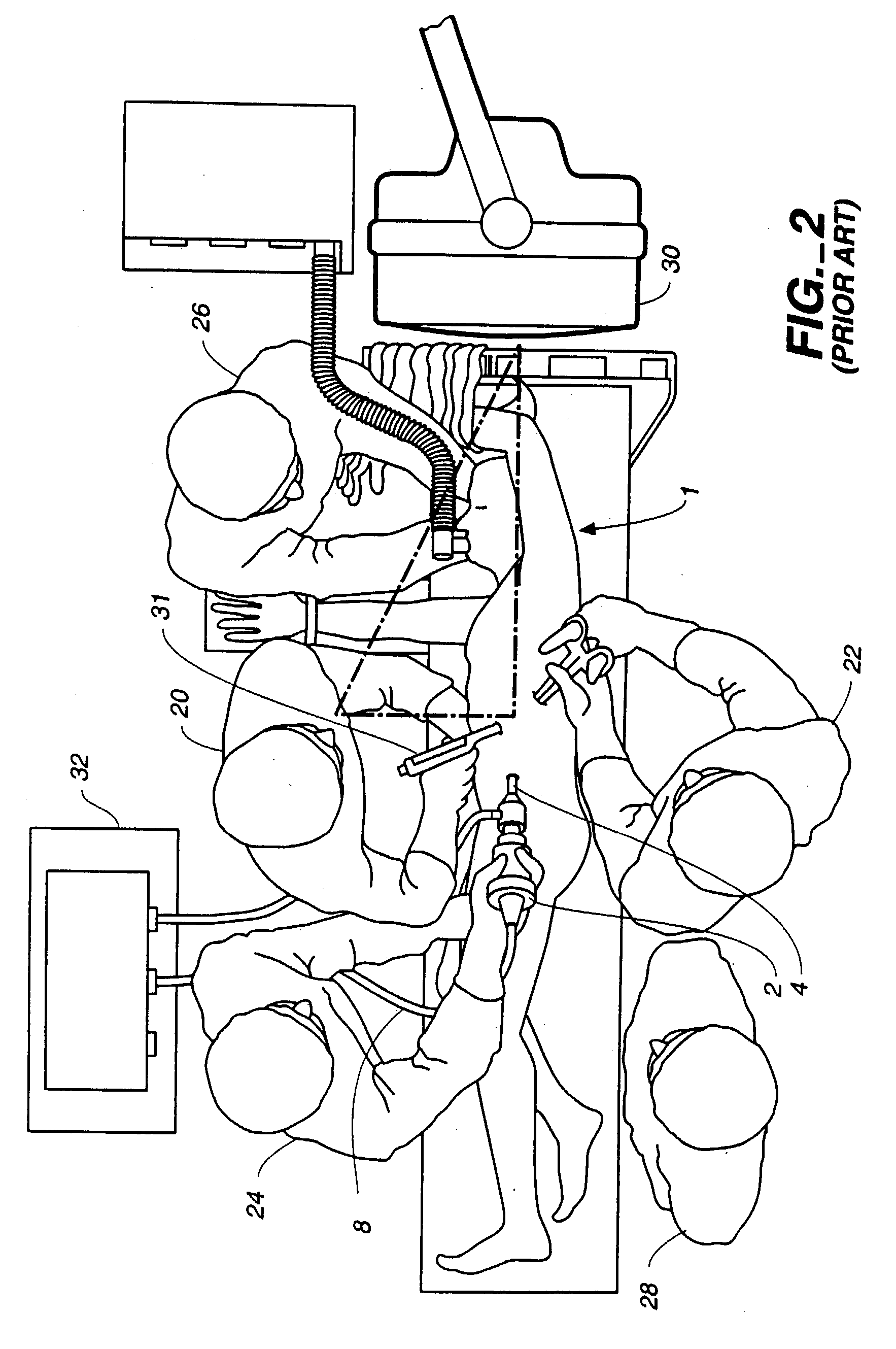
This makes it difficult and uncomfortable for the surgeon to perform the work as he is viewing the screen either a significant distance away from the surgical field or at an angle which is different than he is used to viewing the surgery were it open chest surgery.
In addition, having only a two-dimensional viewing monitor makes the surgery process, particularly suturing, difficult to accomplish.

Method used


Image

  • Visualization during closed-chest surgery
  • Visualization during closed-chest surgery
  • Visualization during closed-chest surgery

Examples


Embodiment Construction

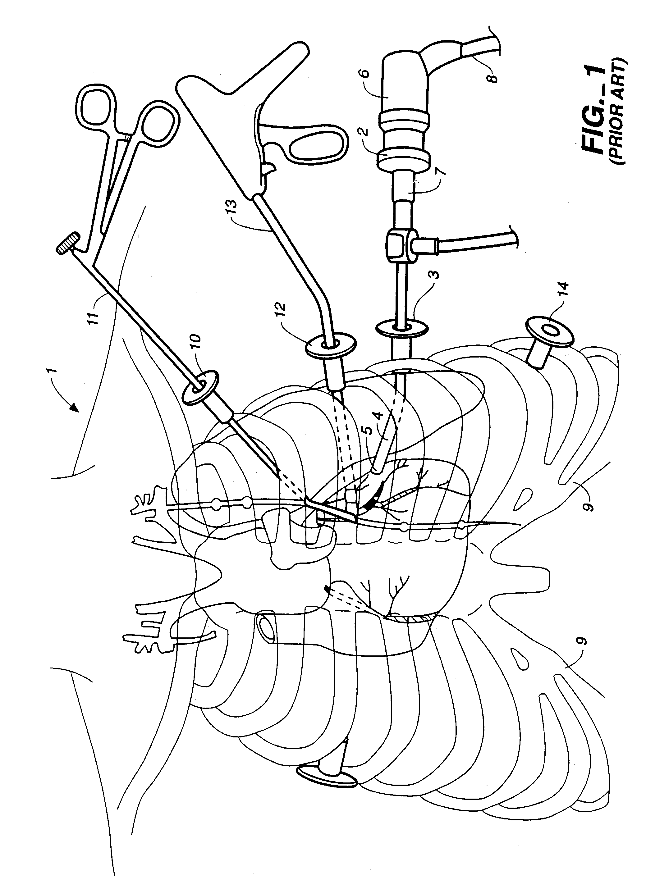
[0058] One aspect of this invention can be considered an improvement of a method for video-assisted, closed-chest diagnostic or surgical treatment of a patient, particularly for cardiac surgery. The improvement is to an existing method that requires the use of a viewing scope (e.g. a thoracoscope or an endoscope) to carry out a surgical procedure. A viewing scope comprises a miniature video camera with a lens means, a light source and transmission means, a processing unit for forming an image and a transmission means for transmitting the image to a viewing monitor. The lens means and light transmission means is usually in a nosepiece that is inserted through a percutaneous hole in the appropriate position of the patient and positioned so that the lens gathers the image that ultimately is transmitted to the viewing monitor. Suitable viewing scopes include those made by Olympus, Solos, Starz, Medical Dynamics, Feijinan, Wolf, Stryher, Linvatic, ACMI, Pentax, Dyonics and Concept. These...


PUM


Abstract

An improvement in a method for closed-chest, video-assisted diagnostic or surgical treatment of a patient is provided. The improvement comprises draping a video monitor with a transparent sterile surgical drape and positioning the draped monitor within the surgical field so that a surgeon can perform an internal surgical or diagnostic procedure and view it on the monitor to provide improved visual alignment for the surgeon. A flexible, sterile drape for covering the video monitor is disclosed. An apparatus for assisting the surgeon in performing closed-chest, video-assisted surgical or diagnostic treatment of a patient is disclosed which comprises a movable cabinet having an adjustable arm to which is affixed a video monitor that can be extended into the surgical field of an operating room to improve the visualization of the surgery performed by the surgeon. An improved operating table for assisting a surgeon in performing closed-chest, video-assisted surgical or diagnostic treatment of a patient is also disclosed along with endoscopic visualization apparatus and an improved design of a view scope that allows the doctor to get closer to a patient during the operation.

Description

[0001] This is a continuation-in-part of Provisional patent application U.S. 60 / 014,922 filed Apr. 10, 1996 in the name of inventors Arthur Bertolero, Raymond Bertolero and Jerome Riebman. This application is related to concurrently-filed patent applications ESTC-001 / 01WO, ESTC-001 / 02WO, ESTC-001 / 04WO and ESTC-001 / 05US. Each of the above-identified patent applications is incorporated herein by reference.[0002] 1. Technical Field[0003] This invention relates methods and apparatus for improving a surgeon's ability to perform video-assisted, closed-chest diagnostic and surgical procedures on patients requiring such procedures. It particularly relates to bringing a viewing monitor into the surgical field and positioning the monitor to provide the surgeon with nearly perfect visual alignment so that he or she has the feel of open-chest surgery. This is achieved by using specifically designed surgical drapes for the viewing monitor and apparatus designed to unobtrusively position the view...

Claims


Application Information

Patent Timeline
IPC IPC(8): A61B1/05A61B1/313A61B1/32A61B17/00A61B17/02A61B17/04A61B17/28A61B17/32A61B17/80A61B19/00A61B19/08A61M1/36A61M25/00A61M29/02
CPCA61B1/05A61M1/3659A61B1/32A61B17/00234A61B17/0206A61B17/2812A61B17/29A61B17/320016A61B17/8085A61B19/081A61B19/26A61B19/52A61B19/5225A61B2017/00199A61B2017/00243A61B2017/0046A61B2017/2904A61B2017/2905A61B2017/2946A61M25/0026A61M25/1027A61M2025/1052A61M2025/1086A61M2025/1097A61M2210/127A61B1/313A61B90/36A61B90/50A61B46/10A61B90/37
Inventor BERTOLERO, ARTHUR A.BERTOLERO, RAYMOND S.RIEBMAN, JEROME B.
Owner ATRICURE
Learn More