
Method and apparatus for knowledge based diagnostic imaging

a knowledge-based diagnostic imaging and knowledge-based technology, applied in the field of medical diagnostic imaging systems, can solve the problems of not every hospital being able to justify the expense of equipment and staff/operators, and the diagnostic imaging system is quite specialized and expensiv

Inactive Publication Date: 2005-01-13
GE MEDICAL SYST GLOBAL TECH CO LLC
View PDF9 Cites 138 Cited by
  • Summary
  • Abstract
  • Description
  • Claims
  • Application Information

AI Technical Summary

Benefits of technology

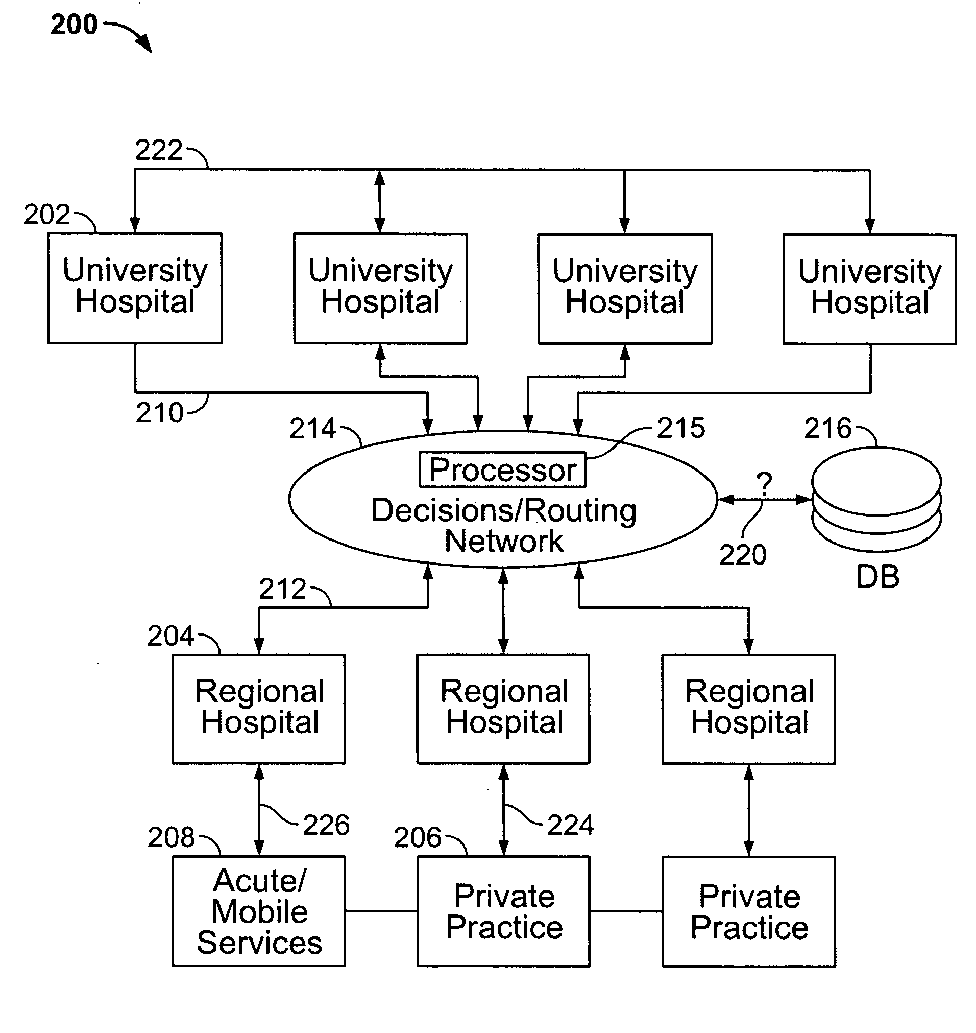
[0006] Certain embodiments of the present invention are directed to knowledge-based diagnostic methods and apparatus that afford a new approach to primary, healthcare (HC) workflow for new patients. The first HC provider that examines each patient is able to utilize diagnostic imaging equipment to provide a more qualified initial diagnosis of the patient. In one applicatio

Problems solved by technology

These diagnostic imaging systems are quite specialized and may be quite expensive.
Due to the nature of each system, technicians, physicians and operators typically expend a significant amount of time in learning how to operate the equipment and interpret images obtained with the equipment.
Hence, not every hospital is able to justify the expense associated with the equipment and the staff / operators that use the equipment.
Also, even when a hospital offers the imaging equipment, the hospital may be unable to justify multiple staff or physicians who are specially trained to utilize the equipment.
This limitation in resources often creates a bottleneck for the use of the equipment and patients are not able to receive immediate examination with such equipment.
Primary healthcare providers today do not utilize diagnostic imaging equipment as part of their normal examination process.
This is due in part to a lack of familiarity and training with such equipment.
Consequently, primary healthcare providers are unable to apply diagnostic imaging in their diagnosis and examinations.
Heretofore, unless the primary healthcare provider has received the particular specialized training needed to utilize diagnostic equipment, the existing healthcare system was unable to provide adequate quality assurance that the primary healthcare provider would properly diagnose a given pathology when viewing the diagnostic images.
There has been no mechanism to educate or share knowledge with the primary healthcare providers that would facilitate such quality assurance.
Existing systems have been unable to provide sufficiently objective and accurate imaging methodologies to support the use of diagnostic imaging equipment by non-specialists.

Method used


Image

  • Method and apparatus for knowledge based diagnostic imaging
  • Method and apparatus for knowledge based diagnostic imaging
  • Method and apparatus for knowledge based diagnostic imaging

Examples


Embodiment Construction

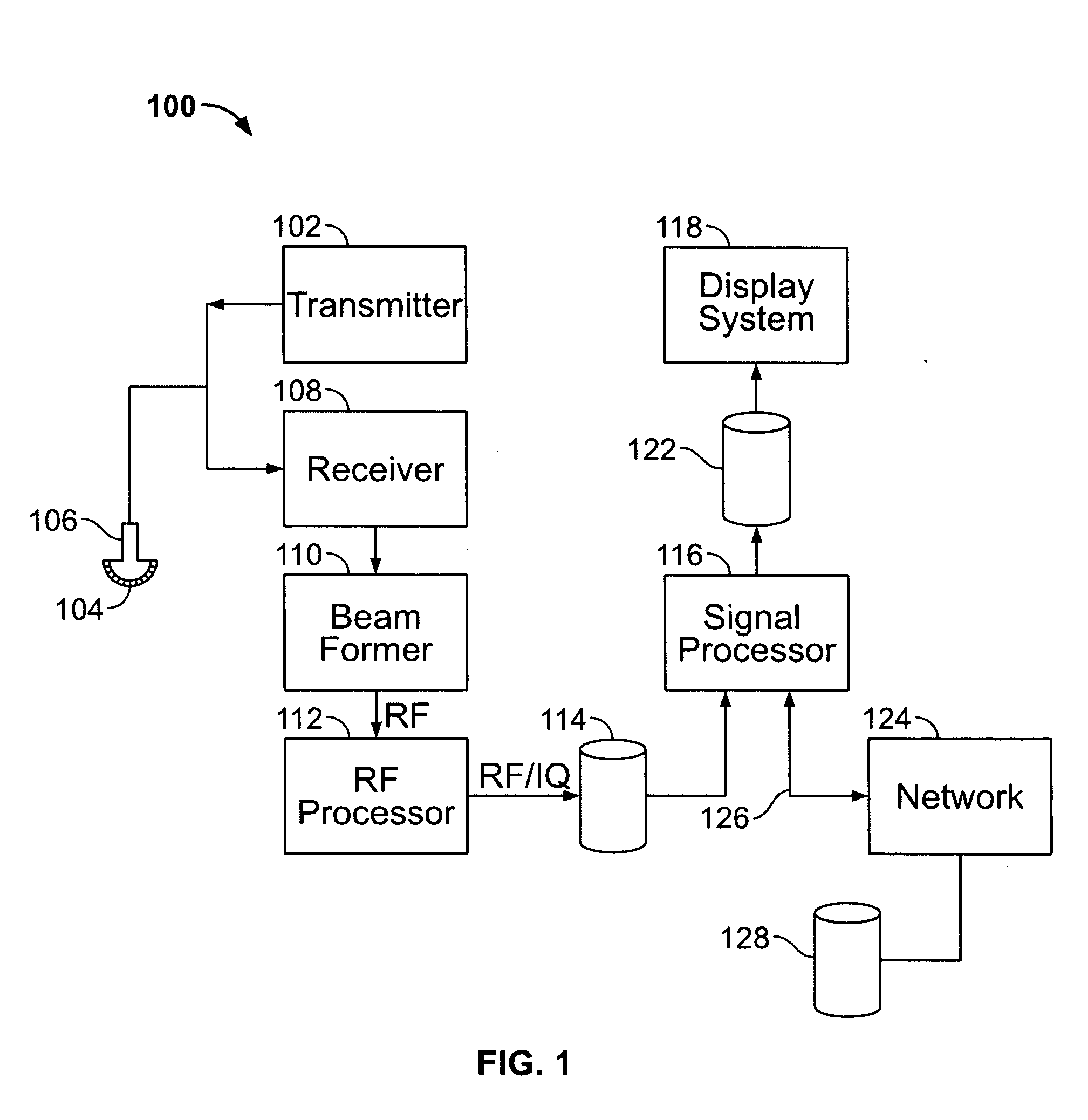
[0014]FIG. 1 illustrates a block diagram of an ultrasound system 100 formed in accordance with an embodiment of the present invention. The ultrasound system 100 includes a transmitter 102 which drives transducers 104 within a probe 106 to emit pulsed signals that are back-scattered from structures in the body, like blood cells or muscular tissue, to produce echoes which return to the transducers 104. The echoes are received by a receiver 108. The received echoes are passed through a beamformer 110, which performs beamforming and outputs an RF signal. The RF signal then passes through an RF processor 112. Alternatively, the RF processor 112 may include a complex demodulator (not shown) that demodulates the RF signal to form IQ data pairs representative of the echo signals. The RF signal or IQ data pairs may then be routed directly to RF / IQ buffer 114 for temporary storage.

[0015] The ultrasound system 100 also includes a signal processor 116 to process the acquired ultrasound informa...


PUM


Abstract

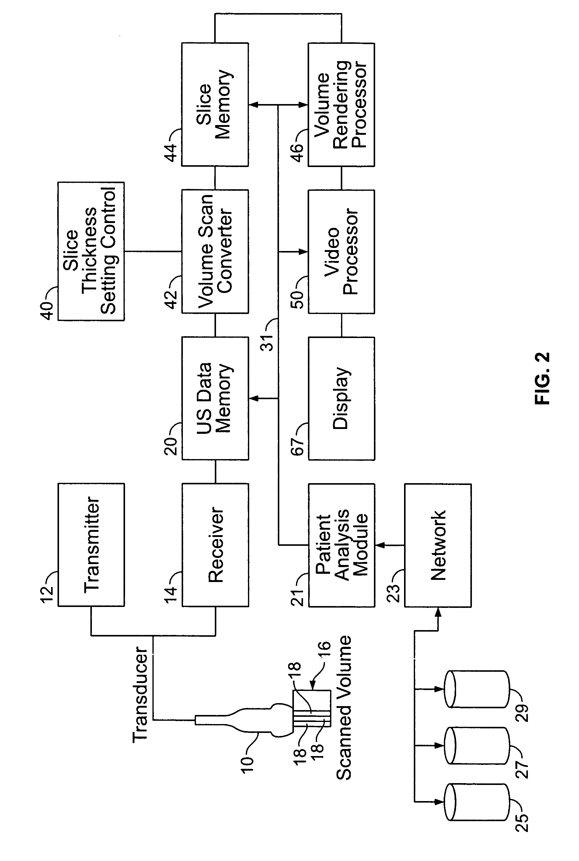
A knowledge based diagnostic imaging system, comprising diagnostic equipment for analyzing a patient to obtain a new patient data set containing at least one of MR data, CT data, ultrasound data, x-ray data, SPECT data and PET data. The diagnostic equipment automatically analyzes the new patient data set with respect to a physiologic parameter of the patient to obtain a patient value for said physiologic parameter. A database containing past patient data sets for previously analyzed patients. The past patient data sets contain data indicative of the physiologic parameter with respect to previously analyzed patients. A network interconnects the diagnostic equipment and the database to support access to the past patient data sets.

Description

RELATED APPLICATION [0001] The present application relates to and claims priority from Provisional Application Ser. No. 60 / 462,012, filed Apr. 11, 2003, titled “Method and Apparatus for Knowledge Based Diagnostic Imaging”, the complete subject matter of which is hereby expressly incorporated in its entirety.BACKGROUND OF THE INVENTION [0002] Today a wide variety of medical diagnostic imaging systems are offered to assist physicians in detecting and diagnosing pathologies. Examples of modalities that offer such diagnostic systems include ultrasound, CT, MR, PET, SPECT and x-ray, as well as mammography and the like. These diagnostic imaging systems are quite specialized and may be quite expensive. Due to the nature of each system, technicians, physicians and operators typically expend a significant amount of time in learning how to operate the equipment and interpret images obtained with the equipment. Specialists may operate the equipment or interpret the resulting images. Hence, not...

Claims


Application Information

Patent Timeline
IPC IPC(8): A61B5/00A61B5/055A61B6/00A61B8/00A61B8/08G16H30/20G16H40/67
CPCA61B5/0002A61B5/055A61B6/00A61B6/541A61B6/56A61B6/563A61B8/483A61B8/08A61B8/543A61B8/56A61B8/565G06F19/321G06F19/3418A61B8/00G16H40/67G16H30/20
Inventor FRIGSTAD, SIGMUNDOLSTAD, BJORN
Owner GE MEDICAL SYST GLOBAL TECH CO LLC
Learn More