

Liquid tissue preparation from histopathologically processed biological samples, tissues and cells
a technology of biological samples and liquid tissues, which is applied in the direction of dissolving, chemical/physical processes, peptides, etc., can solve the problems of lack of sensitivity, inability to quantitatively provide, and limited number of assays that can be performed on formalin-fixed samples
- Summary
- Abstract
- Description
- Claims
- Application Information
AI Technical Summary
Benefits of technology
Problems solved by technology
Method used
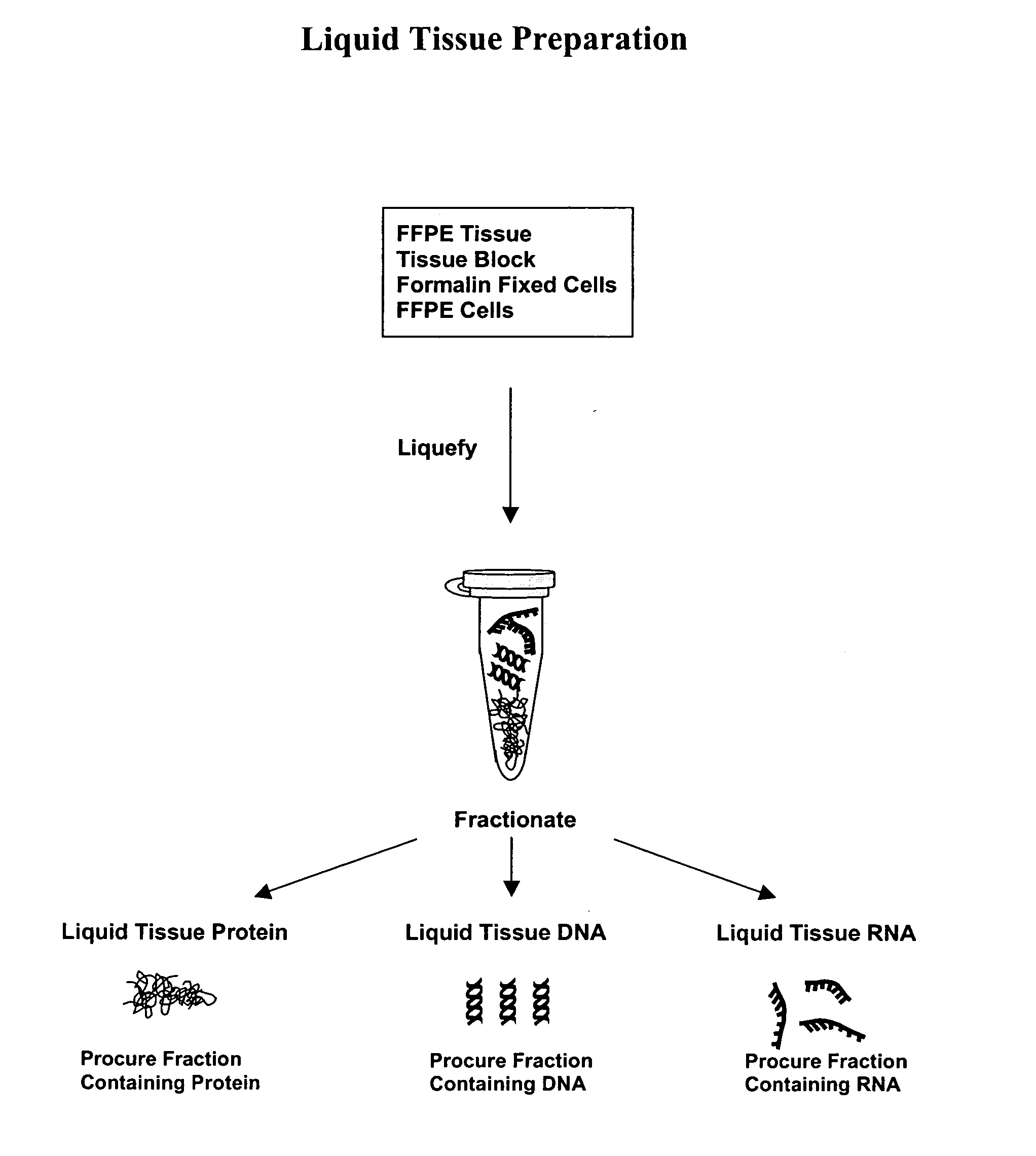
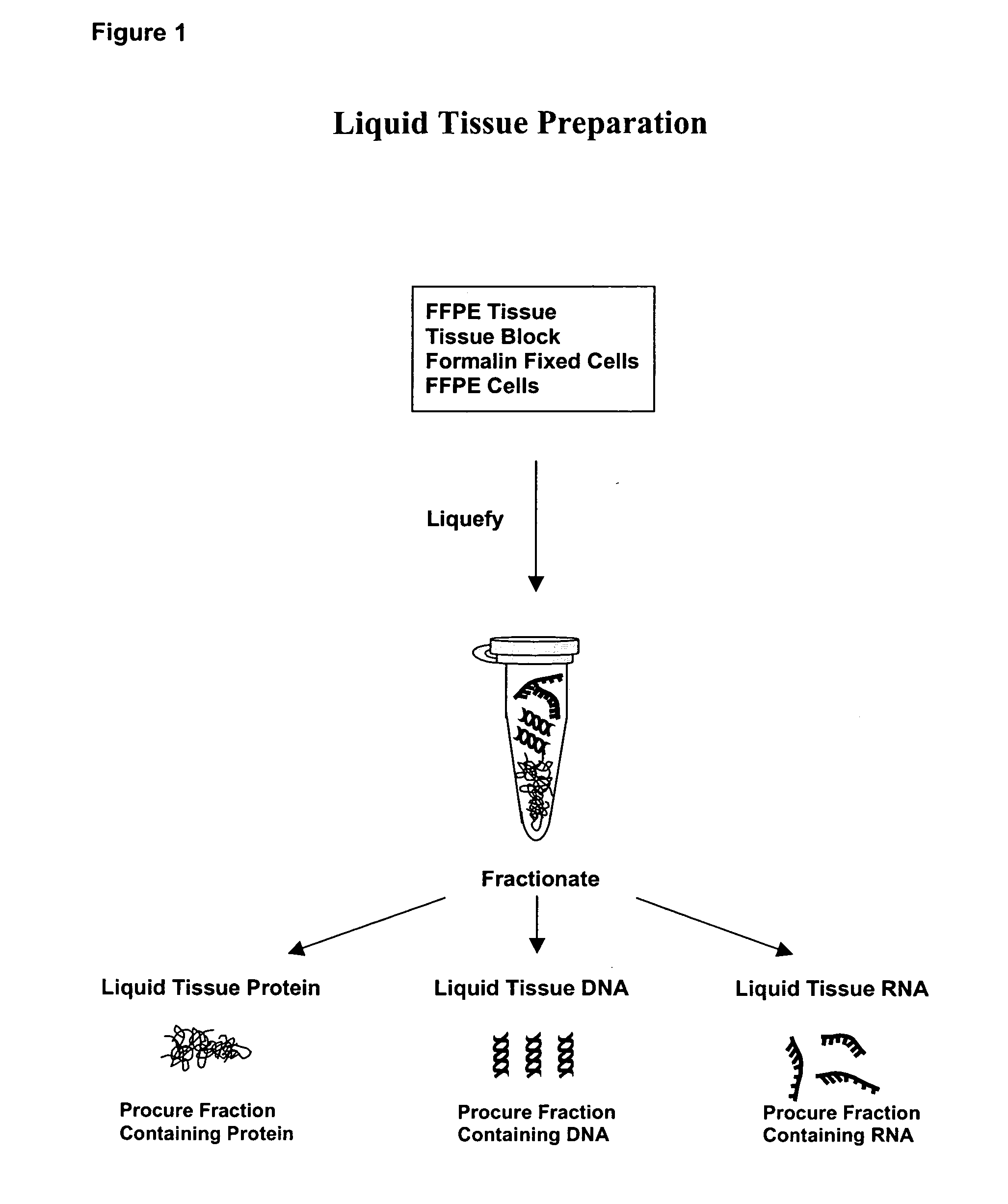
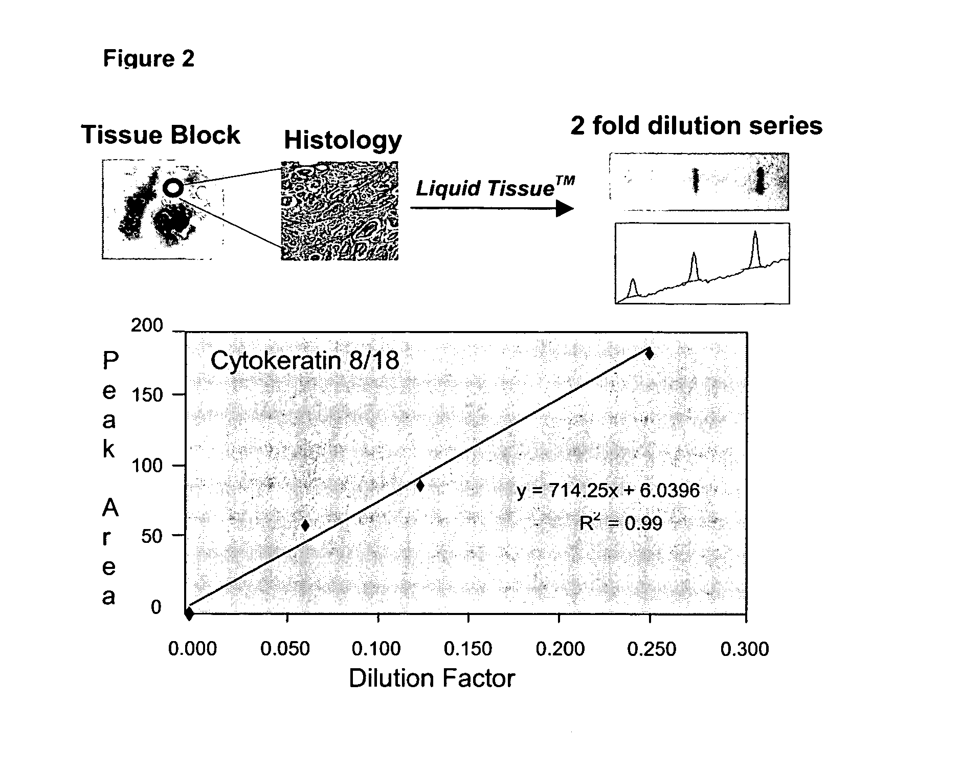
Image

Examples
example 1
Preparation of a Multi-Use Lysate from a Formalin-Fixed Sample
[0075] 1. Place one 2 mm diameter by 25 μm thick section from a tissue punch into a silanized or low protein binding 1.5 ml microcentrifuge tube.
[0076] 2. Add 500 μl of 20 mM Tris-HCl pH 7.8.
[0077] 3. Heat at 95° C. for 1 minute.
[0078] 4. Mix gently on a vortex mixer.
[0079] 5. Carefully, without disturbing the tissue section, remove the buffer using a pipettor.
[0080] 6. Add 750 μl of 20 mM Tris-HCl pH 7.8.
[0081] 7. Heat at 95° C. for 1 minute.
[0082] 8. Carefully, without disturbing the tissue section, remove the buffer using a pipettor.
[0083] 9. Microcentrifuge at 10,000 rpm for 1 minute.
[0084] 10. Remove any residual buffer from the microcentrifuge tube with a pipettor.
[0085] 11. Add 10 μl of reaction buffer (10 mM Tris-HCl pH 7.8, 1.5 mM EDTA, 0.1% Triton X-100, 10% glycerol) to the tube. Make sure that the tissue is at the bottom of the tube and covered with reaction buffer.
[0086] 12. Heat at 95° C. for 1.5...
PUM
| Property | Measurement | Unit |
|---|---|---|
| Temperature | aaaaa | aaaaa |
| Temperature | aaaaa | aaaaa |
| Temperature | aaaaa | aaaaa |
Abstract
Description
Claims
Application Information