Minimally invasive collection probe and methods for the use thereof

a collection probe and minimally invasive technology, applied in the field of medicine, molecular biology and biochemistry, can solve the problems of difficult recognition of certain tumor cells, many challenges, and unreliable and subjective analysis

Active Publication Date: 2022-09-22
BOARD OF RGT THE UNIV OF TEXAS SYST
View PDF11 Cites 0 Cited by
  • Summary
  • Abstract
  • Description
  • Claims
  • Application Information

AI Technical Summary

Benefits of technology

[0070]FIG. 30: Representative mass spectra obtained with the MasSpec Pen from grapes. Results show that the MasSpec Pen analysis was able to produce a spectrum that could be used to characterize the sample.

Problems solved by technology

While intraoperative frozen section analysis has been performed in clinical practice for decades, it presents many challenges.
Freezing artifacts occur during tissue processing and interfere with tissue structure and cell morphology, thus complicating pathologic interpretation, causing the analysis to be unreliable and subjective.
Moreover, certain tumor cells are very difficult to recognize due to their atypical pattern of growth and shape.
However, to date, adequate devices or methodologies have not been developed that provide effective molecular assessment of tissue samples.

Method used


Image

  • Minimally invasive collection probe and methods for the use thereof
  • Minimally invasive collection probe and methods for the use thereof
  • Minimally invasive collection probe and methods for the use thereof

Examples


example 2 -

Example 2-Molecular Profiles and Analysis

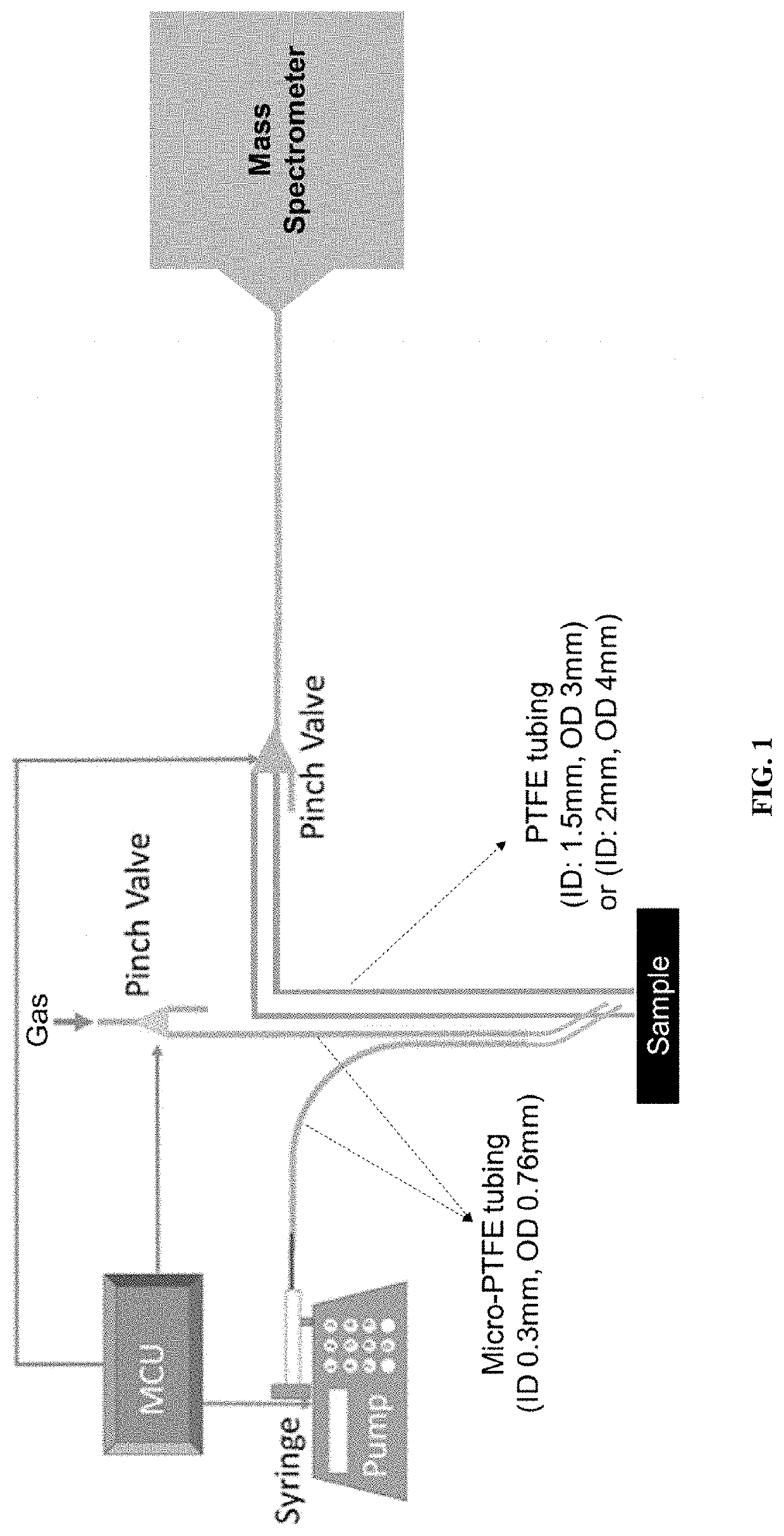
[0107]The system described herein operates by directly connecting the transfer tube to the mass spectrometer inlet for transporting the analyte-containing solvents to the mass spectrometer for molecular analysis. This set up greatly simplifies operational details and precludes the use of ionization sources. After the probe interacts with the tissue, the solvent is then transported to the mass spectrometer and directly infused without the need of an additional ionization source. Since the system is fully automated so that each 10 μL solvent droplet is delivered separately to the inlet, the mass spectrometer operates without any impact on its performance. Rich molecular information is obtained in this manner, similar to what is observed from other solvent-extraction ambient ionization techniques such as desorption electrospray ionization. The ionization mechanism may be similar to inlet ionization. For inlet ionization methods, the ionization o...

example 3 -

Example 3-System Automation for Handheld and Laparoscopic Use

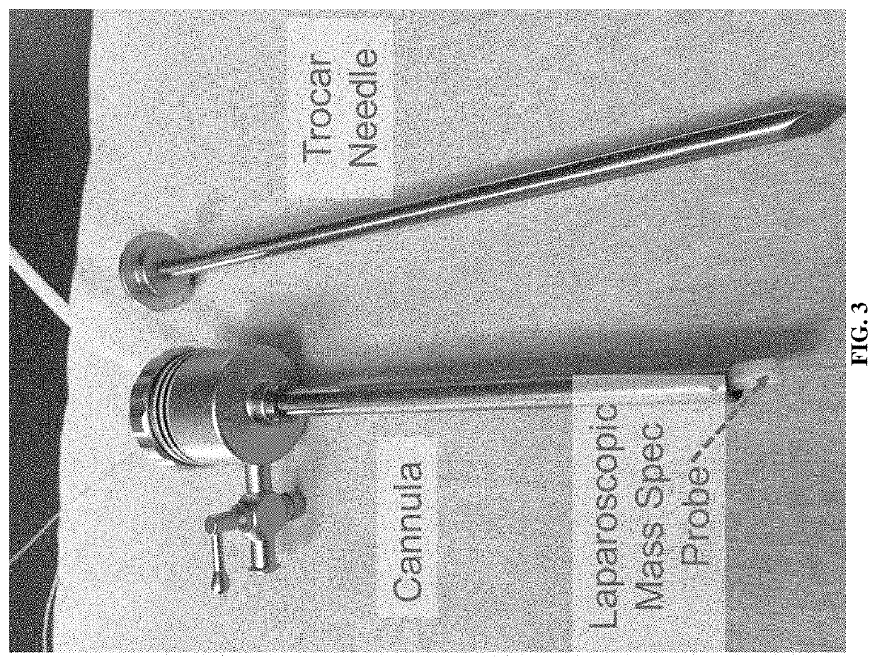
[0118]Because all the materials (PDMS and PTFE) and solvent (only water) used in the minimally invasive probe design are biologically compatible, the system has a high potential to be used in laparoscopic and endoscopic surgeries for real-time analysis. More than that, due to the small dimension of the device, it can be integrated to a robotic surgical system, such as the Da Vinci surgical system, through an accessory port or one of its robotic arms. Several regions of the human body cavity can be quickly sampled during surgery with or without wash / flush steps in between each analysis, and analyzed by using a database of molecular signatures and machine learning algorithms. Therefore, the diagnosing results may be provided in real time for each sampled region. This system can be broadly used in a wide variety of oncological and other surgical interventions (such as endometriosis) for which real-time characterization and di...

example 4 -

Example 4-Materials and Methods

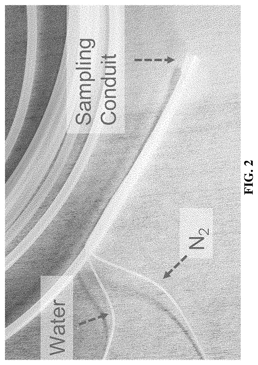
[0132]Laparoscopic MasSpec Pen Design. Three different laparoscopic / robotic MasSpec Pen tips were created from PDMS with diameters of 1.5 mm, 2.7 mm, and 4.0 mm. Grasping fins were built-in unilaterally in order to minimize overall cross section. Two micro-PTFE tubings (OD 0.794 mm, ID 0.339 mm) were grafted to the interior of the PDMS tip near the distal end. The micro-PTFE tubing was terminated 2 millimeters above the reservoir in order to avoid tissue interaction. The PDMS mixing solution from Dow Corning (Midland, Mich.) was molded into negative prints created by a Stratasys uPrint SE Plus 3D Printer (Eden Prairie, Minn., USA). PTFE tubing was purchased from Sigma-Aldrich (St. Louis, Mo., USA), and silicone tubing was purchased from Saint Gobain (Tygon #3550, Malvern, Pa., USA). This design allows the formation of a water droplet at the distal end of the PDMS tip that interacts with tissue to extract cellular lipids and small metabolites via phase ...


PUM

PropertyMeasurementUnit
lengthaaaaaaaaaa
pressureaaaaaaaaaa
pressureaaaaaaaaaa

Abstract

Method and devices are provided for assessing tissue samples from a plurality of tissue sites in a subject using molecular analysis. In certain aspects, devices of the embodiments allow for minimally invasive collection of liquid tissue samples and delivery of the samples for mass spectrometry analysis.

Description

[0001]This application is a continuation of U.S. application Ser. No. 16 / 882,801 filed May 26, 2020, which is a continuation of International Application No. PCT / US2018 / 062625 filed Nov. 27, 2018, which claims the benefit of U.S. Provisional Patent Application Nos. 62 / 591,179, filed Nov. 27, 2017 and No. 62 / 640,385, filed Mar. 8, 2018, each of which are incorporated herein by reference in their entirety.[0002]This invention was made with government support under Grant No. R00CA190783 awarded by the National Institutes of Health. The government has certain rights in the invention.BACKGROUND OF THE INVENTION1. Field of the Invention[0003]The present invention relates generally to the field of medicine, molecular biology and biochemistry. More particularly, it concerns methods and devices for assessment of tissue samples using mass spectrometry.2. Description of Related Art[0004]Clinical diagnosis is commonly performed through the evaluation of tissue samples pre-operatively, intra-ope...

Claims


Application Information

Patent Timeline
Patent Type & Authority Applications(United States)
IPC IPC(8): A61B5/00A61B34/30A61B1/313A61B10/02A61B17/34A61M25/00G01N33/483H01J49/04H01J49/16
CPCA61B5/0075A61B34/30A61B1/3132A61B10/02A61B17/34A61M25/0026G01N33/4833H01J49/0431H01J49/165A61B2090/395G01N1/4055A61B10/0283A61B17/29A61B10/04H01J49/0404H01J49/0459H01J49/04A61B2090/3983A61B10/06
Inventor EBERLIN, LIVIA SCHIAVINATOMILNER, THOMASZHANG, JIALINGGIESE, NOAHKATTA, NITESH
Owner BOARD OF RGT THE UNIV OF TEXAS SYST
Why Eureka
Try Eureka