

Universal collection medium
a collection medium and universal technology, applied in the field of cytological and molecular assays, can solve the problems of difficult or impossible quantitative analysis upon storage, excessive dna, rna and other assayable biomolecules, and less than ideal molecular testing
- Summary
- Abstract
- Description
- Claims
- Application Information
AI Technical Summary
Benefits of technology
Problems solved by technology
Method used
Image

Examples
example 1
General Methods for Nucleic Acid Analysis
[0130]The assay for nucleic acids follows in general principle the method for detecting HIV RNA by the Digene Hybrid Capture HIV Test, described in WO 93 / 10263 by Digene. Briefly, following lysis, 50 μl of probe mix (containing DNA biotinylated probe) was added to each well. The plate was sealed and incubated at 65° C. for 1.5 hours for hybridization to occur. After hybridization, samples were transferred to a strepavidin-coated microplate, and 25 μL of anti-hybrid antibody was added to each well. The plate was agitated at 1100 RPM, for 1 hour, at room temperature. Wells were washed 6X times with 65° C. wash buffer, followed by one wash using distilled water. 100 μl of a chemiluminescent substrate was added to each well and the plate was incubated at room temperature for 30 minutes. The plate was then read in the DML 2000 luminometer. The data was then expressed as signal-to-noise. Using a calibration curve, the chemiluminescent signal genera...
example 2
General Methods for Morphological Analysis
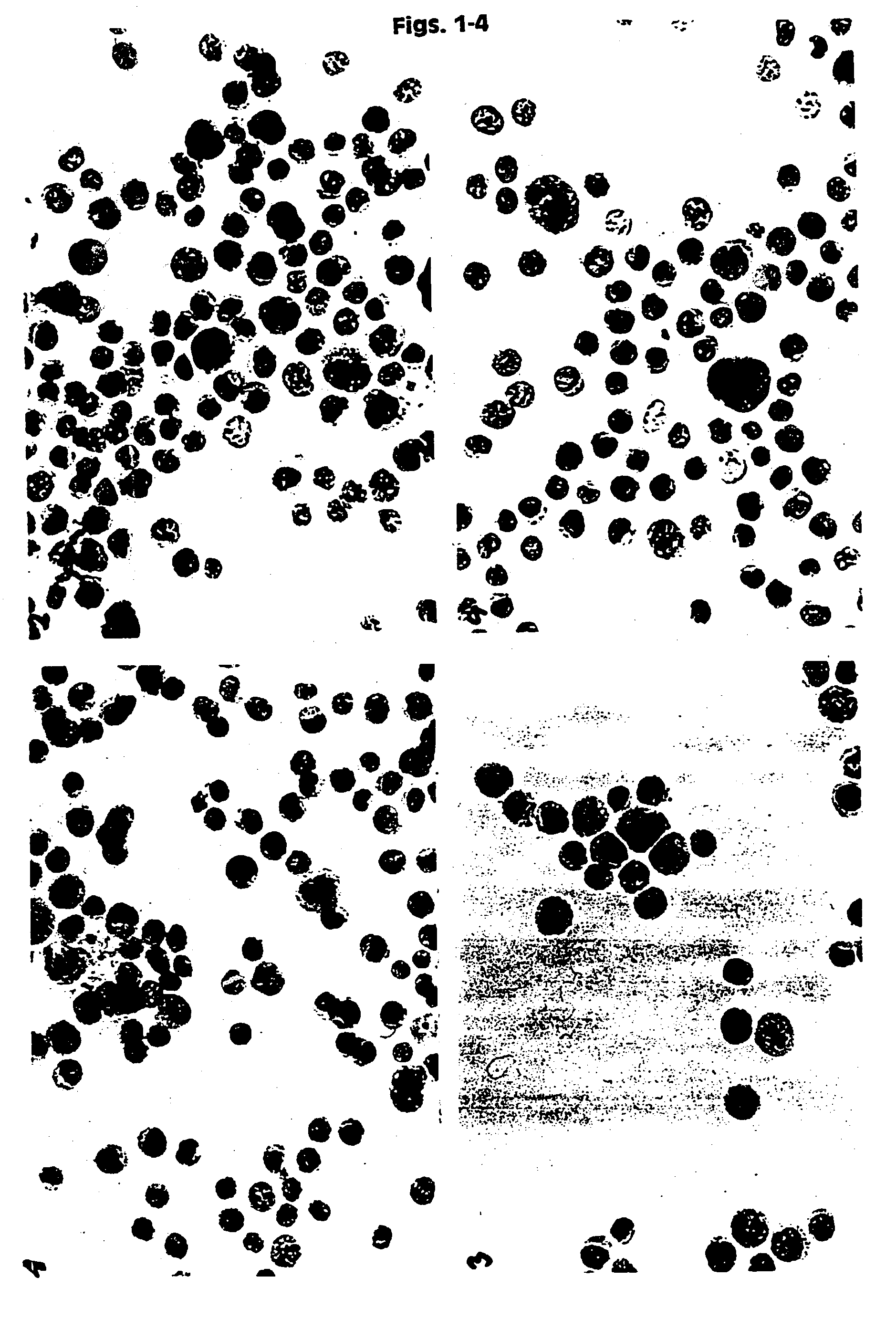
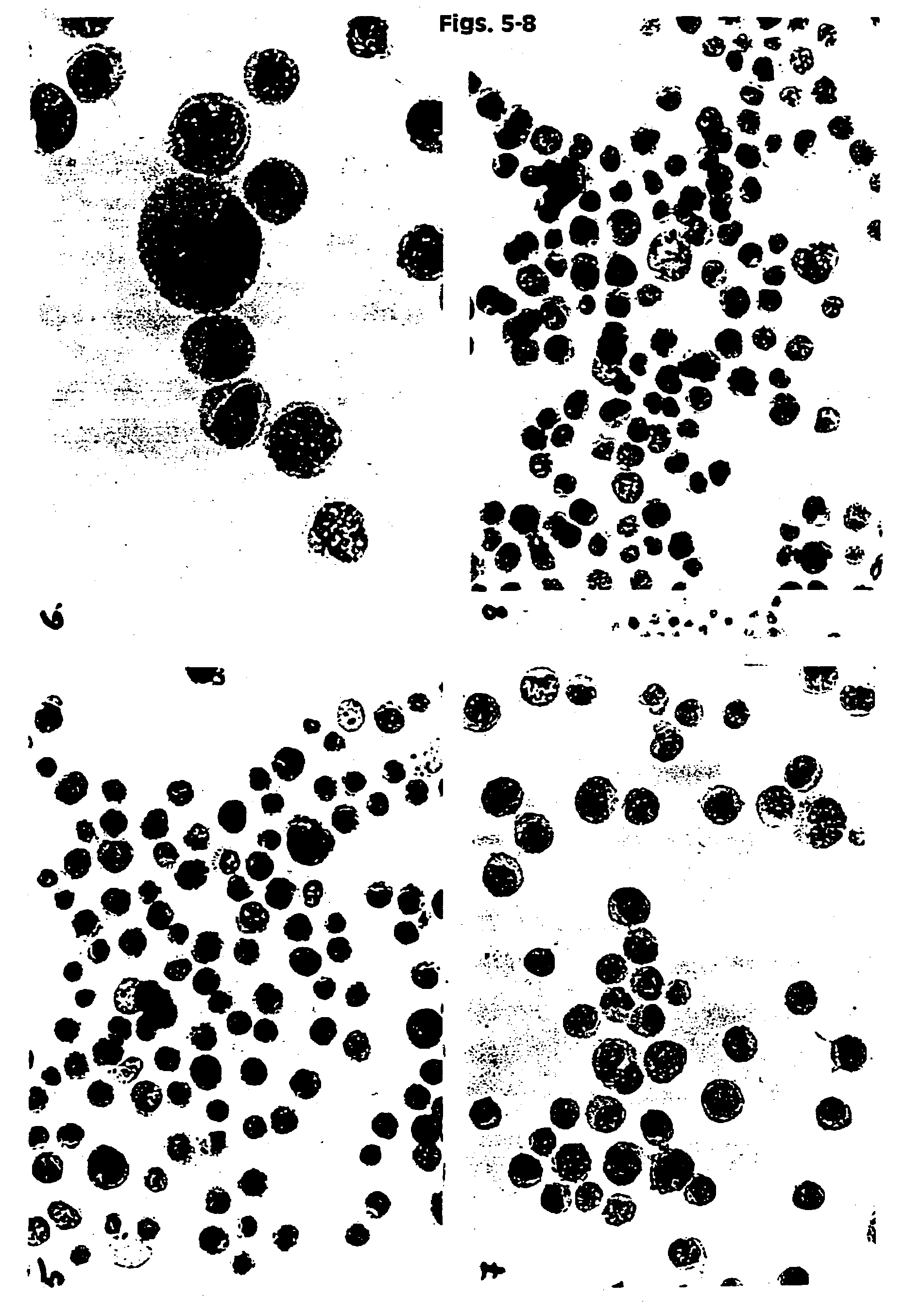
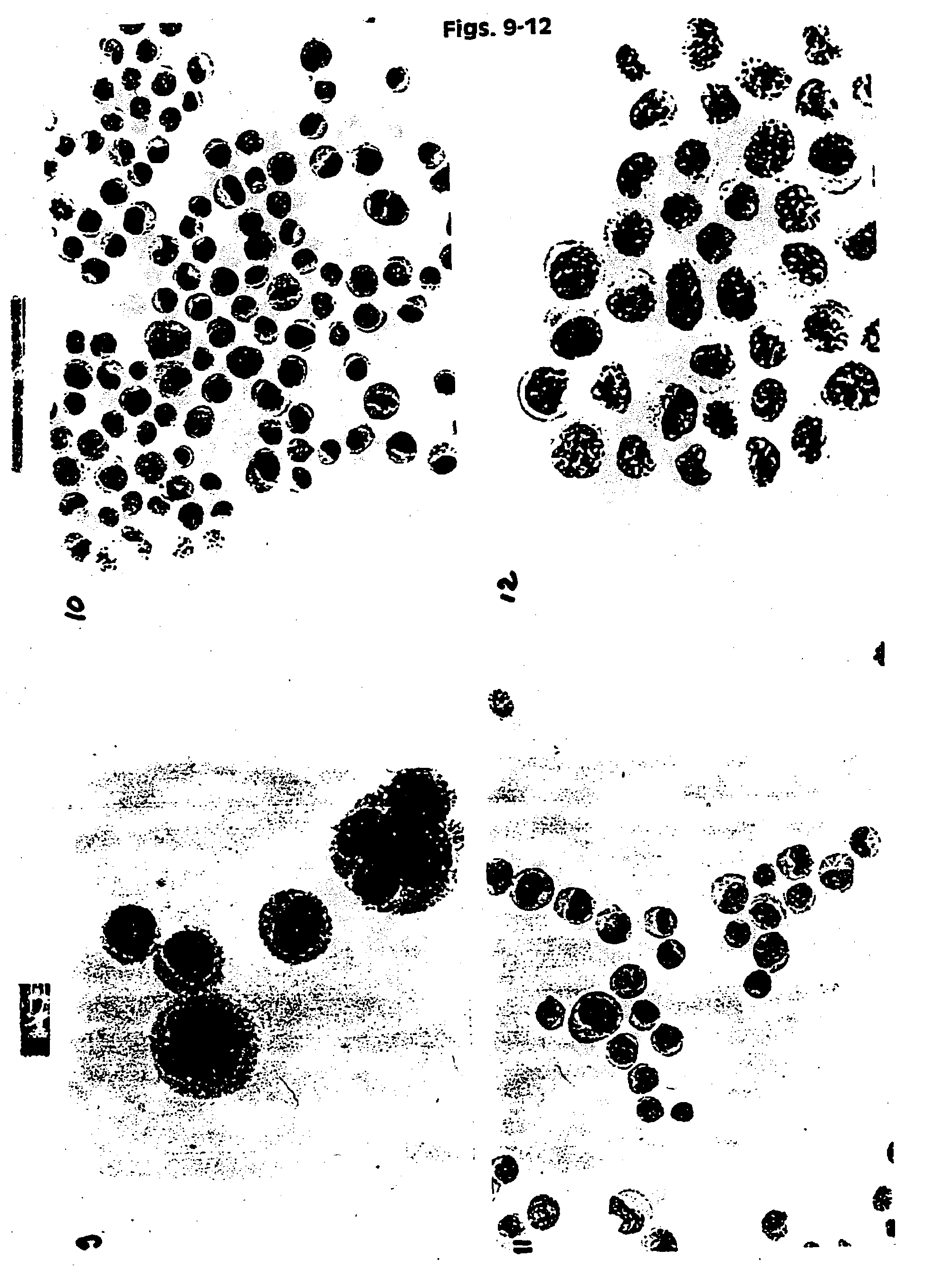
[0131]HPV 16 positive cancer cells (CaSki) were placed in UCM 127, 128, 141, 130, 149 and in two commercially available fixatives PreservCyt (Cytyc Corp.) and CytoRich (AutoCyte). The samples were then stored at ambient temperature. The baseline slides were prepared after 12 hours of storage. Then slides were prepared after 3 and 6 weeks. In addition slides were prepared from UCM 141 and 149 after 6 weeks at RT. The slides were prepared by spotting 200 μl of cell suspension onto polycarbonate filter. The filter was then placed on the glass slide and blotted. The filter was then removed and the slides fixed in 95% ethanol for 5 minutes. The slides were stained using routine Papanicolaou staining and Hematoxylin and Eosin (H&E). The slides were evaluated under a light microscope using different magnifications, and documentation in the form of color pictures was prepared. Additionally a smaller study was performed using a mixture of normal huma...
example 3
HC II HPV DNA Assay Results
[0134]UCM formulations 127, 128, 130 and the STM™ (Digene) control were tested using the Hybrid Capture II HPV DNA Test. A standard HC II HPV Test kit (Digene catalog number 5101-1096) was used. Each collection medium (1 mL) was spiked with 0.8×10⁶ CaSki cells (˜500 copies / cell). This concentration of CaSki cells was chosen because an adequate clinical specimen usually contains about 1×10⁶ cells. The same stock of CaSki cells was then used for morphology study and DNA and RNA testing. A standard volume of 50 μl was used per assay as described in the Package Insert, without any sample preparation modification. A similar STM sample was prepared by spiking the same number of CaSki cells into 1 mL of Digene Sample Transport Medium (STM—this medium is the current medium used for HPV testing. It preserves DNA and RNA but not cell morphology). The HC II HPV test was performed at day “0” (baseline) and after one and six weeks of storage at room temperature. Table ...
PUM
| Property | Measurement | Unit |
|---|---|---|
| Fraction | aaaaa | aaaaa |
| Fraction | aaaaa | aaaaa |
| Fraction | aaaaa | aaaaa |
Abstract
Description
Claims
Application Information