Compositions for isolating a cDNA encoding a membrane-bound protein

a membrane-bound protein and cdna technology, applied in the field of selective and efficient isolating genes encoding membrane-bound proteins, can solve the problems of not being able to know whether it is a secretory protein or a membrane-bound protein, and not being able to achieve the effect of superior selectivity

Inactive Publication Date: 2005-01-27
CHUGAI PHARMA CO LTD
View PDF2 Cites 0 Cited by
  • Summary
  • Abstract
  • Description
  • Claims
  • Application Information

AI Technical Summary

Benefits of technology

[0010] The present invention solves the problems of the TMT method and provides a gene cloning method with a superior selectivity.
[0011] A feature of the present invention is to isolate a gene encoding a membrane-bound protein by linking a functional protein to the fusion protein itself, differing from the conventional TMT method that carries an epitope recognizing an antibody. The present method thus enabled the selective isolation of genes encoding membrane-bound proteins.
[0040] In order to increase the cloning efficiency in the invention, for example, when using single-chain Fv as the functional protein, it is preferable that the C terminus contains a small amount of hydrophobic amino acids, and specifically, a single-chain Fv in which the elbow region has been deleted as described in Examples below can be used. Also, it is preferable that, in the present invention, stability and expression efficiency can be increased by ligating further a domain of secretory protein origin, for example, a DNA encoding amino acids of the constant region of an antibody described in Examples below, to the C terminus of single-chain Fv.
[0049] The obtained cDNA is ligated to a vector. At this instance, cDNA can be introduced into the vector by introducing it downstream of the 3′ side of a functional protein encoding-DNA that is already contained in the vector. For this purpose, a suitable restriction enzyme site, for example, a multi-cloning site is designed downstream of the 3′ side of the DNA encoding the functional protein, and the cDNA is introduced into that site. Also, cDNA may be ligated first downstream of the DNA encoding the functional protein, and then the obtained DNA may be introduced into the vector. The DNA construct can be introduced into a suitable restriction enzyme site comprised in a vector DNA. When preparing the vector, the DNA encoding the functional protein and the cDNA located downstream of the 3′ side may be directly ligated, or may be ligated via a DNA encoding a peptide linker to enable easy binding of the functional protein to the antigen.

Problems solved by technology

However, the identification of a protein having a desired function from these ESTs is by no means an easy task, and in order to predict and analyze the function of an encoded-protein from gene sequence information, a great deal of time and efforts are required.
However, even if a gene fragment encoding a protein comprising a signal sequence is obtained by this method, one cannot know whether it is a secretory protein, or whether it is a membrane-bound protein.
Also, this method requires the utilization of a cDNA library comprising a 5′ end, but techniques for efficiently constructing a cDNA library that selectively contains a 5′ end are not necessarily easy, versatile techniques.

Method used


Image

  • Compositions for isolating a cDNA encoding a membrane-bound protein
  • Compositions for isolating a cDNA encoding a membrane-bound protein
  • Compositions for isolating a cDNA encoding a membrane-bound protein

Examples


example 1

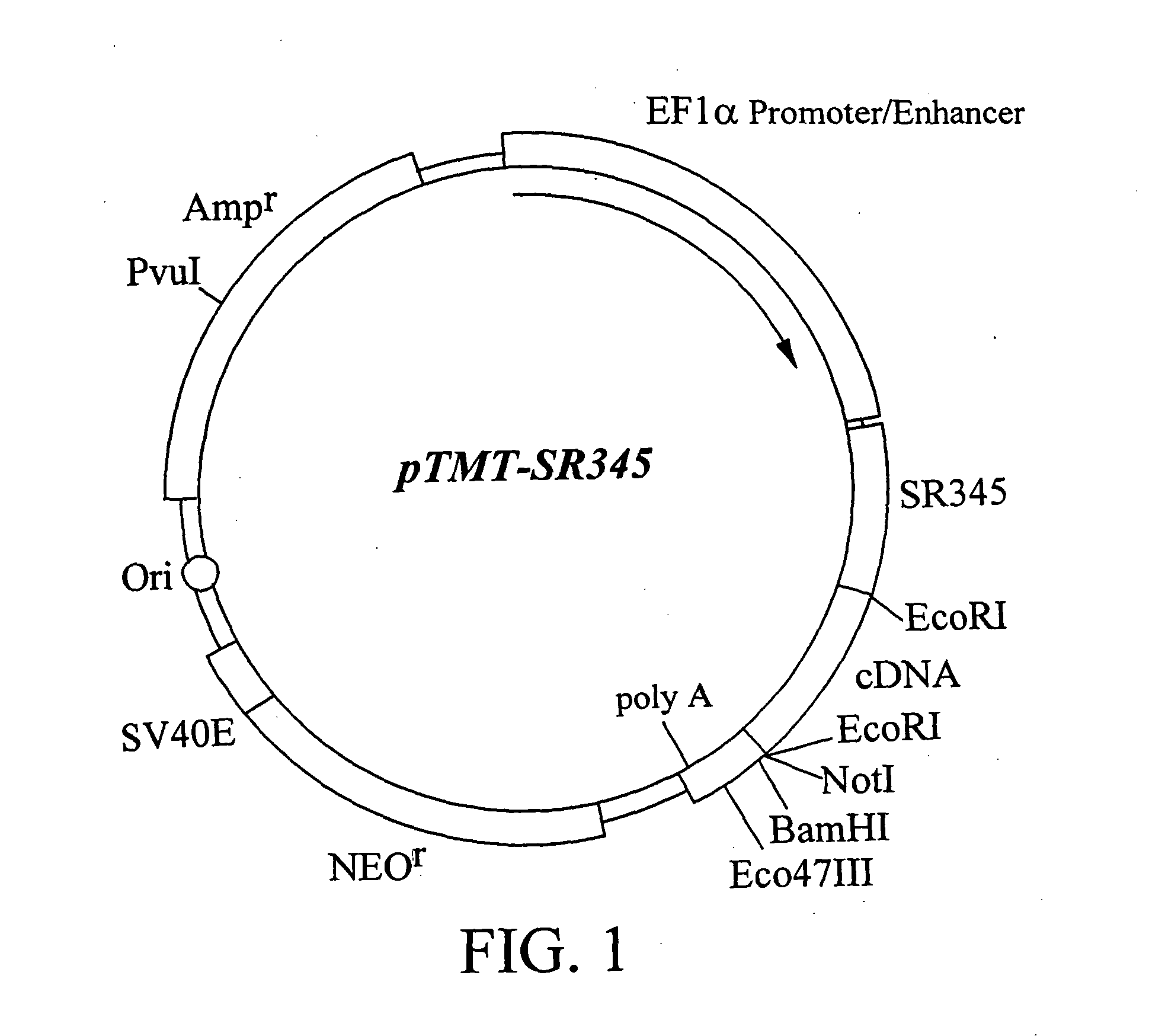
Construction of Expression Cloning Vector pTMT-SR345

[0069] Expression cloning vector pTMT-SR345 was constructed. SR345, encoded by the DNA contained in expression cloning vector pTMT-SR345, is the extracellular region portion of human IL-6 receptor, and consists of 345 amino acid residues from the N terminus. In the expression cloning vector pTMT-SR345, the protein encoded by cDNA inserted downstream of the DNA encoding SR345 is expressed as a fusion protein with SR345. The nucleotide sequence of SR345 is shown in SEQ ID NO: 2 together with the amino acid sequence.

[0070] First, in order to amplify the app. 1.1 kb fragment containing the cDNA encoding SR345 from the cDNA of IL-6 receptor (Yamasaki, K. et al, Science (1988) 241, 825-828), PCR primers IL6R1 (SEQ ID NO: 3) and IL6R2 (SEQ ID NO: 4) were designed. A PCR reaction mixture (100 ml) containing 10 mM Tris-HCl (pH8.3), 50 mM KCl, 0.1 mM dNTPs, 1.5 mM MgCl2, 100 pmol each of the above-mentioned primers, 100 ng of template DNA ...

example 2

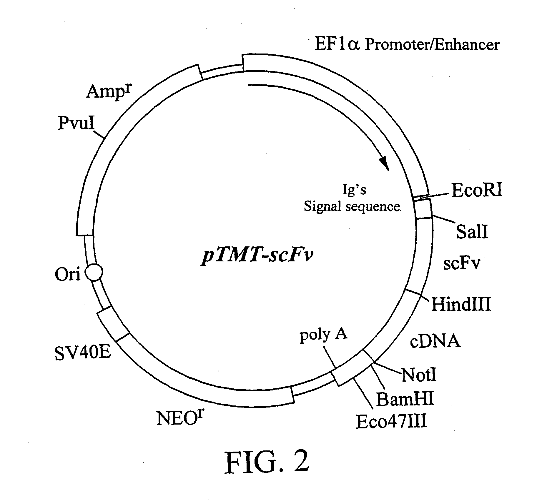
Construction of Expression Vector pTMT-scFv

[0072] Expression vector pTMT-scFv was constructed. The single-chain antibody (scFv) encoded by the DNA contained in the expression vector pTMT-scFv was designed using the variable region of the humanized monoclonal antibody PM-1, which recognizes human IL-6 receptor, and a linker region. In the expression vector pTMT-scFv, the protein encoded by the cDNA inserted downstream of the DNA encoding scFv, is expressed as a fusion protein with scFv. The nucleotide sequence of scFv gene is shown in SEQ ID NO: 5 together with the amino acid sequence.

[0073] 1) Amplification of the DNA Fragment Encoding Antibody V Region

[0074] The genes of humanized PM1 antibody H chain and L chain V region (Sato, K et al, Cancer Res. (1993) 53, 851-856) were amplified by PCR. Backward primer TMT1 (SEQ ID NO: 6) for H chain V region was designed in such a manner that it should hybridize to DNA encoding the N terminus of H chain V region and comprise a SalI restric...

example 3

Construction of SR345-gp130 and scFV-gp130 Fusion Protein Expression Systems

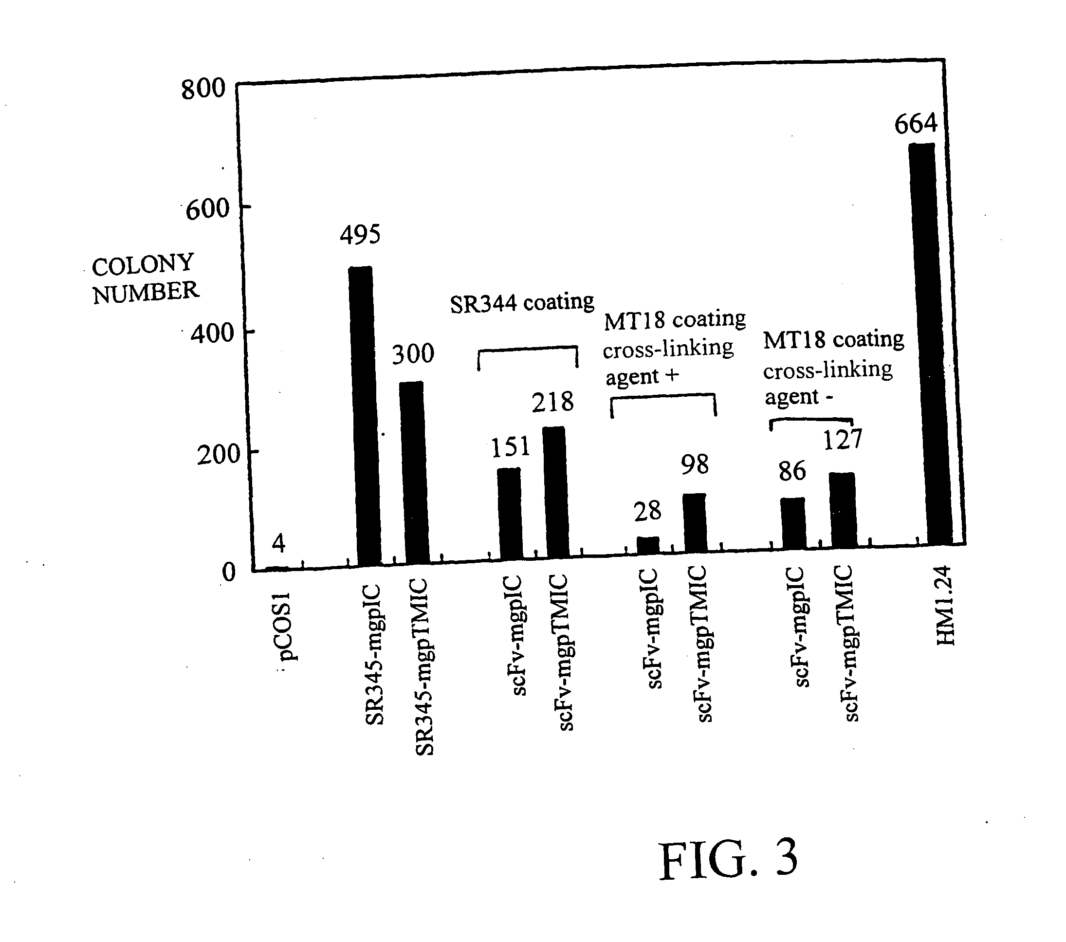
[0081] (A) SR345-gp130

[0082] The cytokine signal transduction molecule gp130 is a type I membrane-bound protein (Taga, T. et al., Cell (1989) 58, 573-581 Saito, M., et al., J. Immunol. (1992) 148, 4066-4071). A portion of mouse gp130 cDNA was ligated downstream of a cDNA encoding soluble-type IL-6 receptor (SR345) of the expression vector pTMT-SR345, to express a fusion protein comprising SR345 and a partial sequence of mouse gp130, in COS cells. Two types of fusion proteins were constructed according to their differences in the gp130 partial regions. One of them is a membrane-bound fusion protein (SR345-mgpTMIC) in which the transmembrane region of gp130 and the subsequent intracellular region are ligated, and the other is a secretory fusion protein (SR345-mgpIC) in which only the intracellular region of gp130 is ligated. SEQ ID NO: 17 shows the amino acid sequence and the nucleotide sequence of full-leng...


PUM

PropertyMeasurementUnit
melting pointaaaaaaaaaa
temperatureaaaaaaaaaa

Abstract

A method for isolating a gene encoding a membrane-bound protein characterized by fusing a functional protein with a fused protein itself, which differs from the existing TMT method in which an epitope recognized by an antibody is carried in a fused protein. This method enabled selective isolation of a gene encoding a membrane-bound protein.

Description

TECHNICAL FIELD [0001] The present invention relates to a novel gene-cloning-method for selectively and efficiently isolating genes encoding membrane-bound proteins. BACKGROUND ART [0002] Proteins synthesized in cells can be categorized by their individual characteristics into those localized in intracellular organelles, such as nucleus, mitochondria, cytoplasm, etc.; those that function by binding to the cell membrane, such as receptors and channeling molecules; and those that function by being secreted to the cell exterior, such as growth factors and cytokines, etc. In particular, protein molecules bound to the cell membrane are responsible for biologically important functions, such as cellular responses towards growth factors and differentiation factors, inflammatory responses, cell-cell interactions, hormone responses, and so on, and therefore, can be target molecules for diagnostic and therapeutic drugs for various types of disorders. [0003] In recent years, as typified by the ...

Claims


Application Information

Patent Timeline
Patent Type & Authority Applications(United States)
IPC IPC(8): C07K14/705C07K16/00C07K16/28C12N15/10C12N15/12C12N15/13
CPCC07K14/705C07K16/00C07K16/2866C12N15/1051C07K2317/622C07K2319/00C12N15/1034C07K2317/24C12N15/10
Inventor TSUCHIYA, MASAYUKISAITO, MIKIYOSHIOHTOMO, TOSHIHIKO
Owner CHUGAI PHARMA CO LTD
Why Eureka
Try Eureka