Transabdominal examination, monitoring and imaging of tissue

a transabdominal or traxsthoracic technology, applied in the field of invivo examination, transabdominal or traxsthoracic noninvasive examination, can solve the problems of system not being very sensitive to tissue oxygenation, path cannot be approximated by a straight line, and infants are particularly at high risk of brain injury

Inactive Publication Date: 2005-02-17
CHANCE BRITTON
View PDF99 Cites 41 Cited by
  • Summary
  • Abstract
  • Description
  • Claims
  • Application Information

AI Technical Summary

Problems solved by technology

However, the visible or IR radiation is strongly scattered and absorbed in biological tissue, and the migration path cannot be approximated by a straight line, making inapplicable certain aspects of cross-sectional imaging techniques.
However, these systems are not very sensitive to tissue oxygenation.
Premature fetuses and infants are particularly at high risk of developing brain injury.
None of the existing diagnostic methods for fe

Method used


Image

  • Transabdominal examination, monitoring and imaging of tissue
  • Transabdominal examination, monitoring and imaging of tissue
  • Transabdominal examination, monitoring and imaging of tissue

Examples


Embodiment Construction

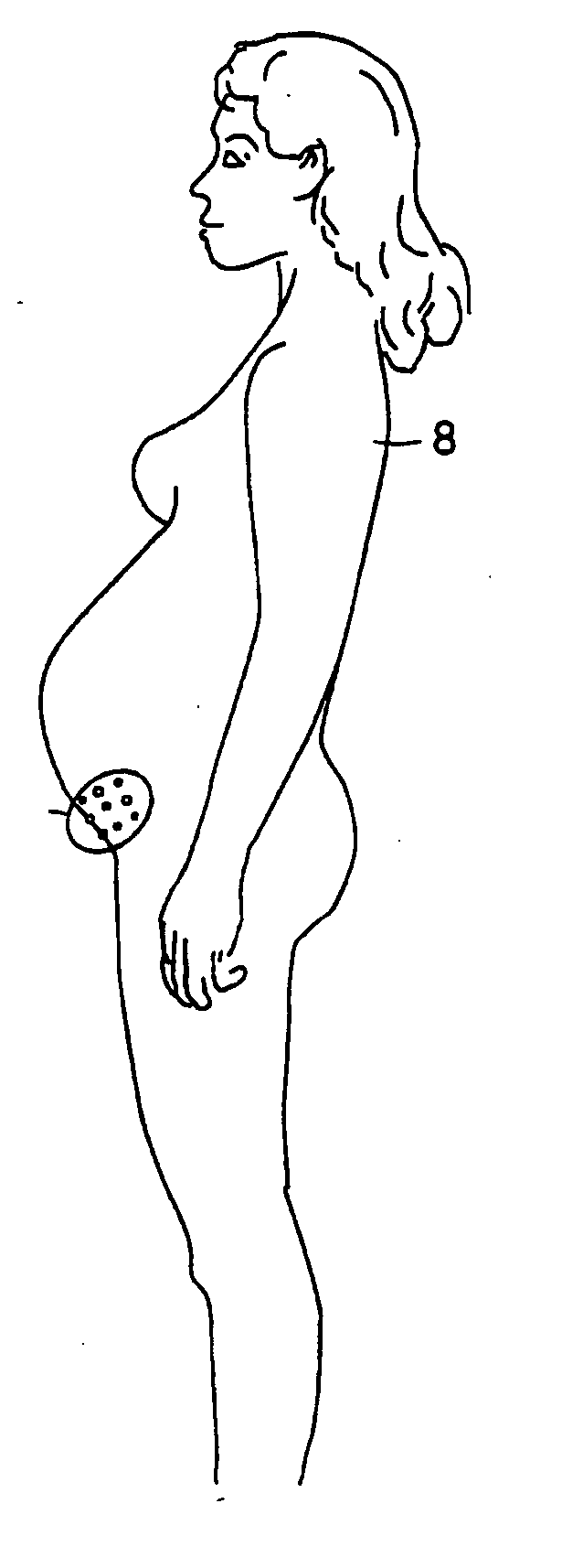
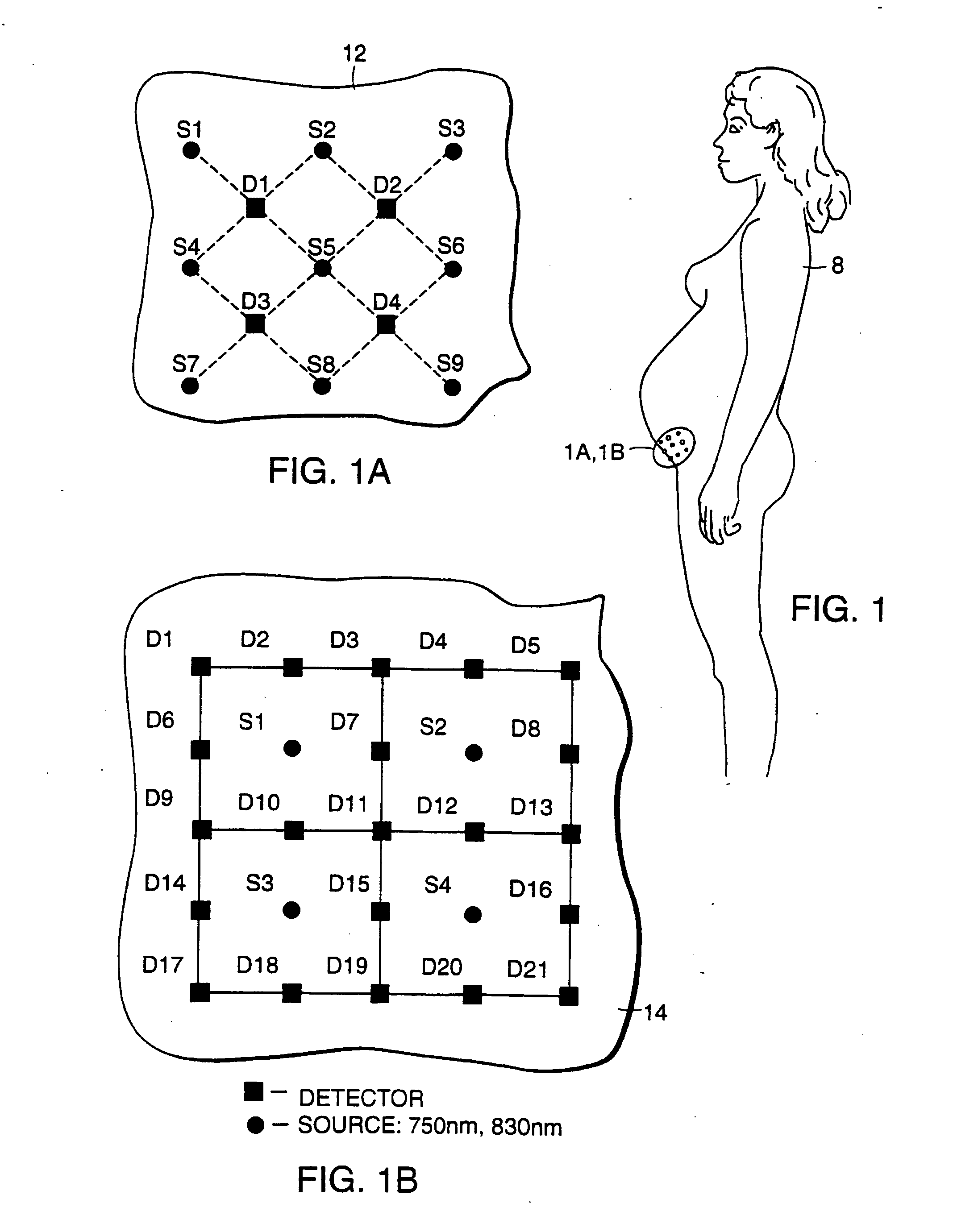
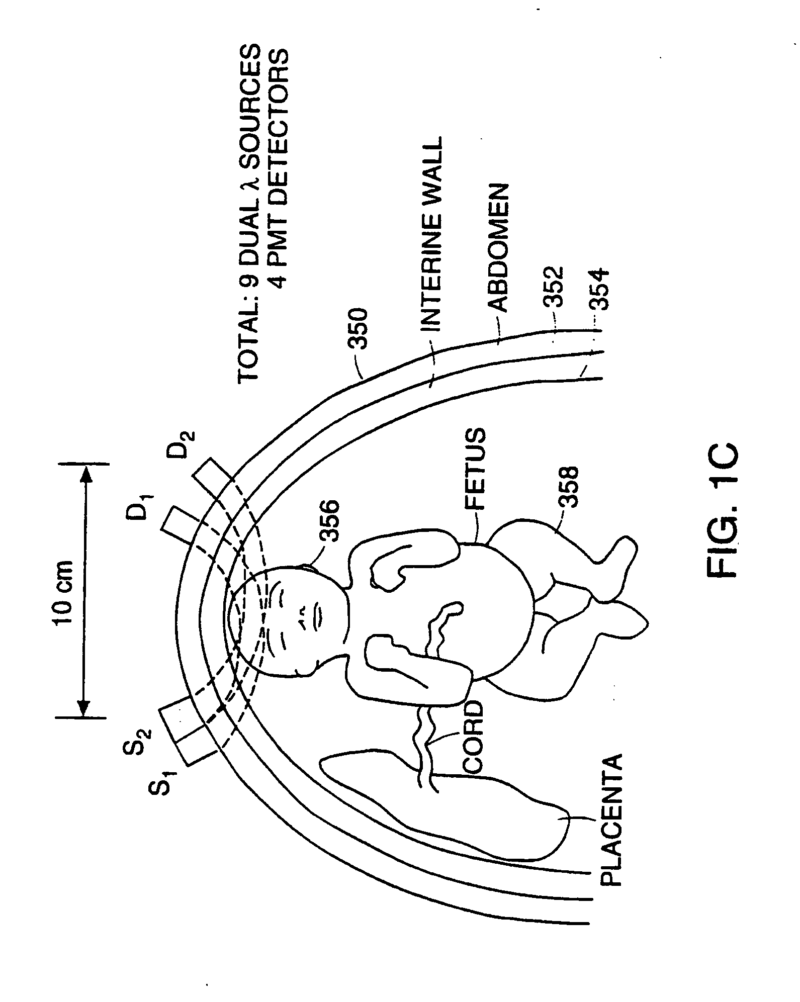
Referring to FIGS. 1 through 2B, a selected fetal tissue of a fetus inside a female subject 8 is examined non-invasively using an imaging system connected to an optical module 12 or 14. Optical modules 12 and 14 include a multiplicity of light sources (e.g., laser diodes, LEDs, flashlight bulbs) providing light in the visible to infrared range and light detectors (e.g., photo multiplier tubes, Si diode detector, PIN, avalanche or other diode detectors), which may also include interference filters. The light sources and the light detectors are arranged to form selected geometrical patterns that provide a multiplicity of source-detector paths of photon migration inside the examined organ. The imaging system provides an in vivo image of the examined tissue. The image shows a location and size of an abnormal structure in the tissue, such as a tumor or bleeding. Furthermore, the image provides a qualitative and quantitative measure (e.g., metabolism, metabolic biochemistry, pathophysiol...


PUM


Abstract

An optical examination technique employs an optical system (15, 45, 100, 150, 200, 260 or 300) for in vivo, non-invasive examination of internal tissue of a subject. The optical system includes an optical module (12 or 14), a controller and a processor. The optical module is arranged for placement on the exterior of the abdomen or chest. The module includes an array of optical input ports and optical detection ports located in a selected geometrical pattern to provide a multiplicity of photon migration paths targeted to examine a selected tissue region, such as an internal organ or an in utero fetus. Each optical input port is constructed to introduce into the examined tissue visible or infrared light emitted from a light source. Each optical detection port is constructed to provide light from the tissue to a light detector. The controller is constructed and arranged to activate one or several light sources and light detectors so that the light detector detects light that has migrated over at least one of the photon migration paths. The processor receives signals corresponding to the detected light and forms at least one data set used for tissue examination.

Description

This application claims priority from U.S. Provisional Application Ser. No. 60 / 074,642 filed on Feb. 13, 1998, U.S. Provisional Application Ser. No. 60 / 098,172 filed on Aug. 26, 1998, and U.S. Provisional Application Ser. No. 60 / 098,018 filed on Aug. 26, 1998, all of which are incorporated by reference as if fully set forth herein. THE FIELD OF THE INVENTION The present invention relates to non-invasive, in-vivo examination, imaging and characrization of biological tissue using visible or infra-red radiation, and more particularly to transabdominal or traxsthoracic non-invasive examination, monitoring and imaging of internal tissue or an in utero fetus. BACKGROUND Traditionally, X-rays or γ-rays has been used to examine and image biological tissue. This radiation propagates in the tissue on straight, ballistic tracks, i.e., scattering of the radiation is negligible. Thus, imaging is based on evaluation of the absorption levels of different tissue types. For example, in roentgenog...

Claims


Application Information

Patent Timeline
IPC IPC(8): A61B5/00A61B10/00A61B5/145A61B5/1455A61B5/1464
CPCA61B5/0059A61B5/0073A61B5/0091A61B5/02411A61B5/06A61B5/14542A61B2562/046A61B5/1464A61B5/4312A61B5/4362A61B5/7285A61B2562/0233A61B2562/0242A61B5/14553A61B2503/02A61B5/0042
Inventor CHANCE, BRITTON
Owner CHANCE BRITTON
Why Eureka
Try Eureka