Medical image generating apparatus and method, and program

a technology of medical image and generating apparatus, applied in the field of medical image generating apparatus, can solve the problems of unclear surface drawing, inability to clearly display the shape of the surface, and inability to display the surface,

Active Publication Date: 2005-03-24
ZIOSOFT
View PDF8 Cites 15 Cited by
  • Summary
  • Abstract
  • Description
  • Claims
  • Application Information

AI Technical Summary

Problems solved by technology

However, in the ray casting using the gradient, though the abnormal region on the surface can be clearly displayed, for example, an abnormal region developed invasively in the interior of tissue cannot be displayed. FIG. 10 shows an example in which an intestinal tract where an invasive abnormal region develops is displayed as a three-dimensional image using the aforementioned method.
However, as recognized by comparison between FIG. 11 and FIG. 10, according to this method, drawing of the surface portion becomes unclear and the shape of the surface is not clearly displayed.
In other words, though the invasive abnormal region is clearly displayed, there occurs such trouble that the polyp and the like on the surface are not displayed.
Namely, complicated working and much time are required, preventing a rapid diagnosis.

Method used


Image

  • Medical image generating apparatus and method, and program
  • Medical image generating apparatus and method, and program
  • Medical image generating apparatus and method, and program

Examples


Embodiment Construction

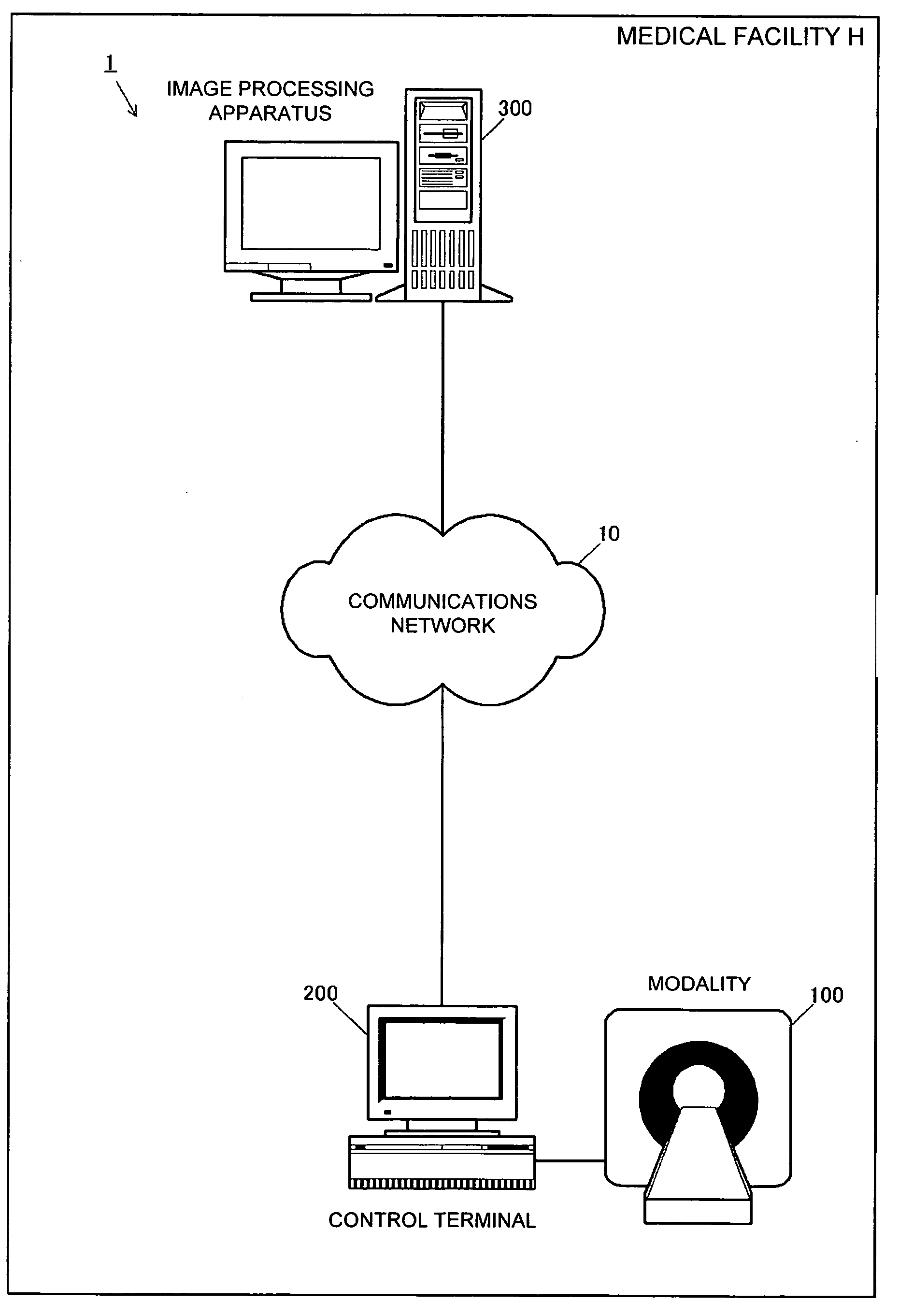
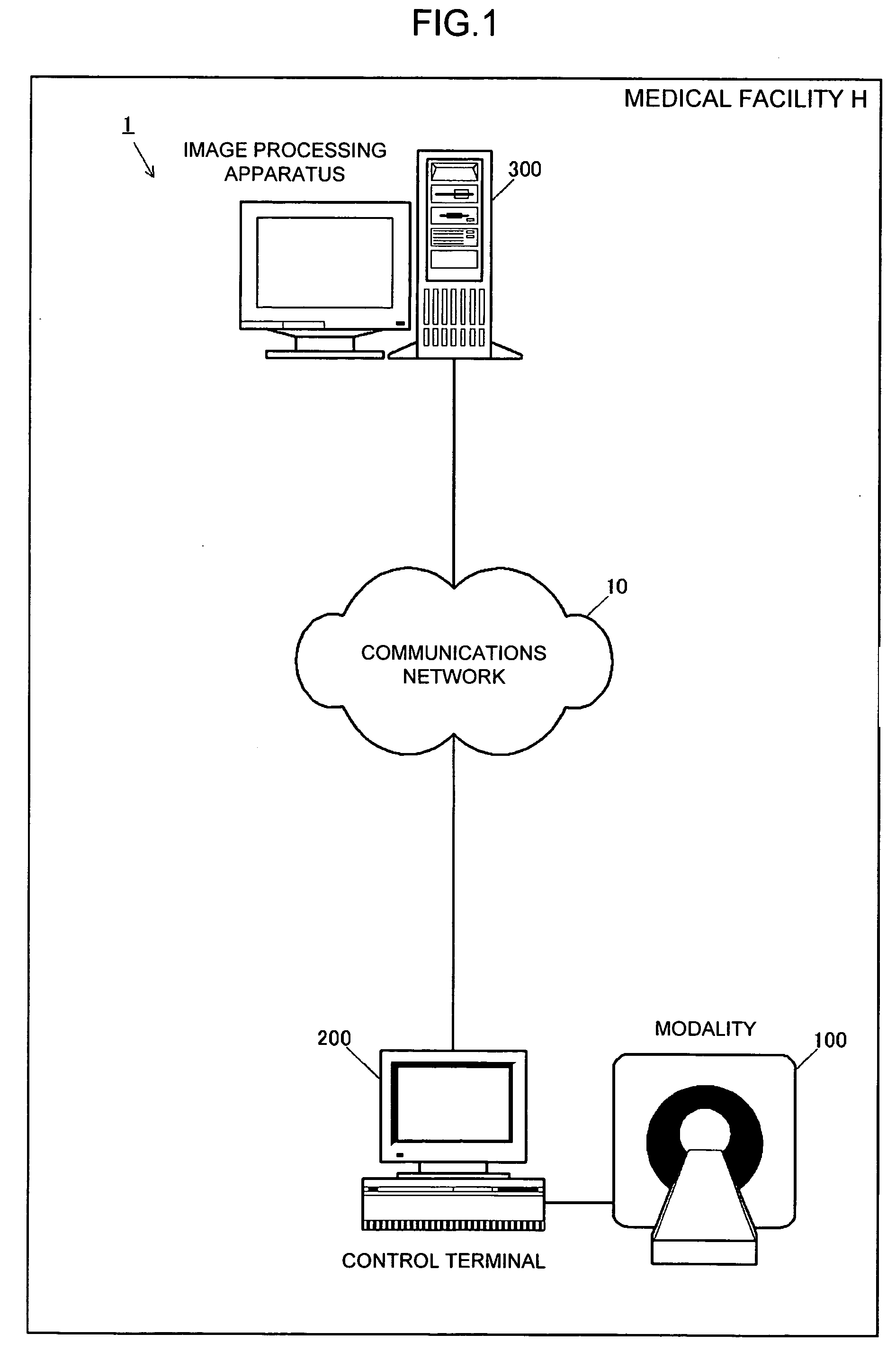
[0030]FIG. 1 is a view schematically illustrating a configuration of an imaging diagnostic system according to an embodiment of the present invention applicable to diagnostic imaging used in medical facilities. As illustrated in the figure, an imaging diagnostic system 1 includes a communications network 10, a modality 100, a control terminal 200, and an image processing apparatus 300.

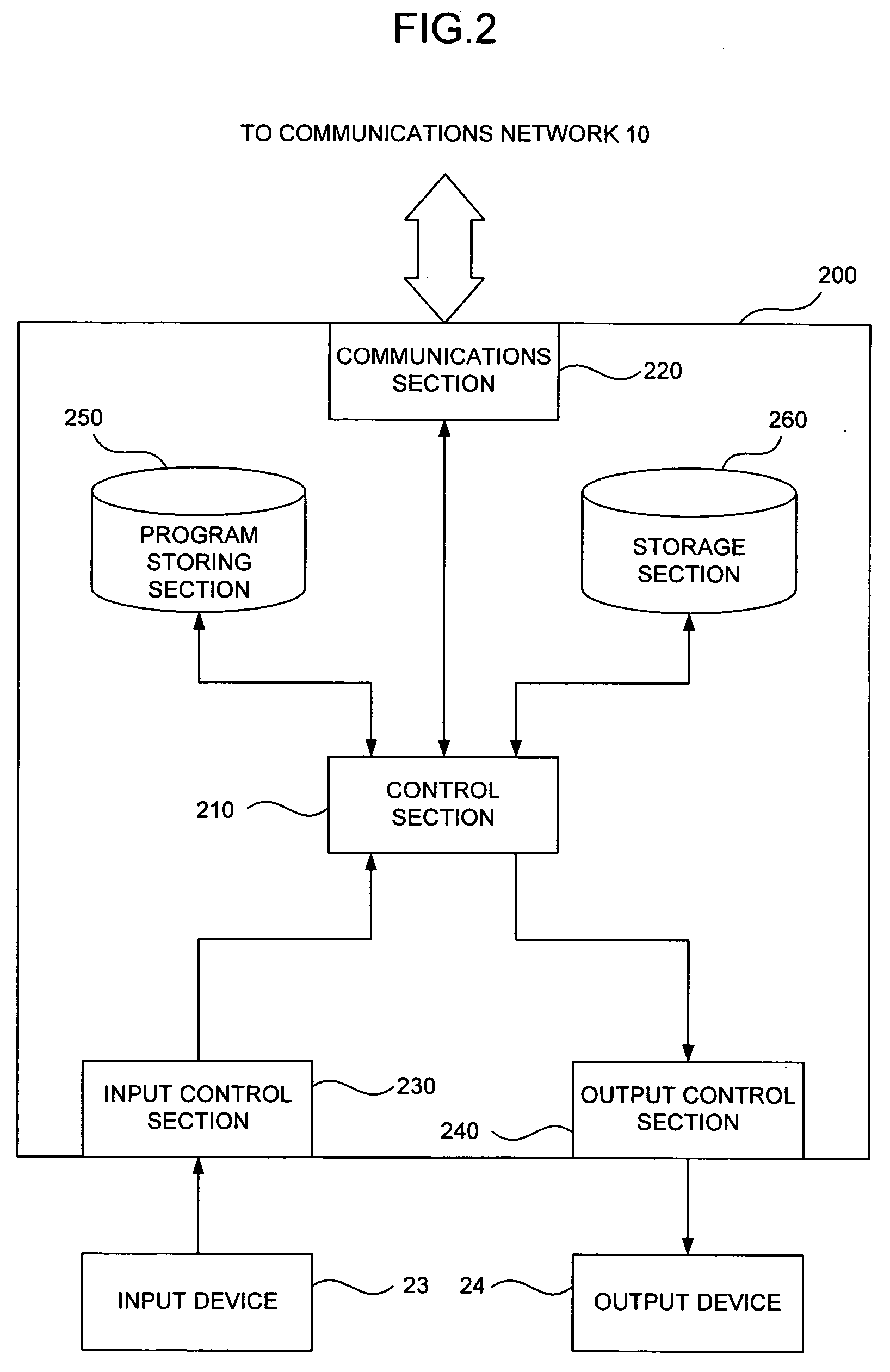
[0031] The communications network 10 is a communications network that connects the control terminal 200 to the image processing apparatus 300 in a medical facility H to carry out information transmission therebetween. The communications network 10 carries out information transmission based on a predetermined communication protocol such as DICOM (Digital Imaging and Communications in Medicine) and the like.

[0032] The modality 100 is an imaging device that images an interior of a human body, and for example, a CT scanner (Computed Tomographic apparatus), a helical CT, an MRI (magnetic resonance imaging...


PUM


Abstract

An image processing apparatus acquires voxel data obtained by imaging an interior of a living body by a modality and generates a three-dimensional medical image by volume rendering using a ray casting method. The image processing apparatus generates the three-dimensional medical image wherein shade of a surface of an inner wall of an intestinal tract is clearly displayed and an abnormal region invasively developed in the interior of the inner wall is distinguishably displayed based on color information corresponding to voxel data placed at a position shifted from the surface by a predetermined distance.

Description

BACKGROUND OF THE INVENTION [0001] 1. Field of the Invention [0002] The present invention relates to a medical image generating apparatus that generates a three-dimensional image used in diagnostic imaging, imaging diagnostic system using the apparatus, medical image generating method and medical image generating program, and particularly to a medical image generating apparatus, which is suitable for diagnosis of an invasive abnormal region, and system, method and program. [0003] 2. Description of the Related Art [0004] Conventionally, there is used imaging diagnostic in which a tomographic image of an interior of a human body is imaged by an imaging device (modality) such as a CT (Computed Tomography) scanner, an MRI (Magnetic Resonance Imaging) and the like and the result is used in diagnosis of disease. Also, such a method is established that a three-dimensional image of such as a predetermined internal organ, an organ and the like is generated from the obtained tomographic image...

Claims


Application Information

Patent Timeline
Patent Type & Authority Applications(United States)
IPC IPC(8): G01R33/32A61B5/00A61B5/05A61B5/055A61B6/03G06T15/08
CPCG06T15/08G06T15/06
Inventor MATSUMOTO, KAZUHIKO
Owner ZIOSOFT
Why Eureka
Try Eureka