Medical image processing apparatus

a technology of medical image and processing apparatus, which is applied in the field of medical image processing apparatus, can solve the problems of complicated conventional approach, and achieve the effect of reducing the screening load of a doctor and improving the accuracy of image measuremen

Inactive Publication Date: 2005-03-31
KONICA MINOLTA MEDICAL & GRAPHICS INC
View PDF7 Cites 22 Cited by
  • Summary
  • Abstract
  • Description
  • Claims
  • Application Information

AI Technical Summary

Benefits of technology

In accordance with the invention, it is possible to improve the accuracy of the image measurement by a doctor, and alleviate the screening load of a doctor.

Problems solved by technology

However, the conventional approach suffers from being too complicated in that it requires a user, according to the individual diagnosis purpose as well as the image usage, to specify the output destination of the medical image and the diagnosis aid information, and the output format.

Method used


Image

  • Medical image processing apparatus
  • Medical image processing apparatus
  • Medical image processing apparatus

Examples


Embodiment Construction

The embodiments of the invention are detailed as follows with reference to the drawings.

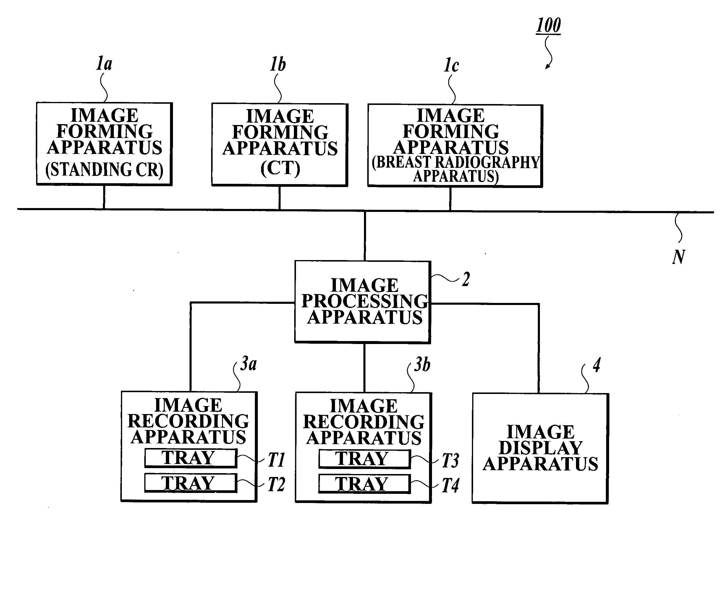
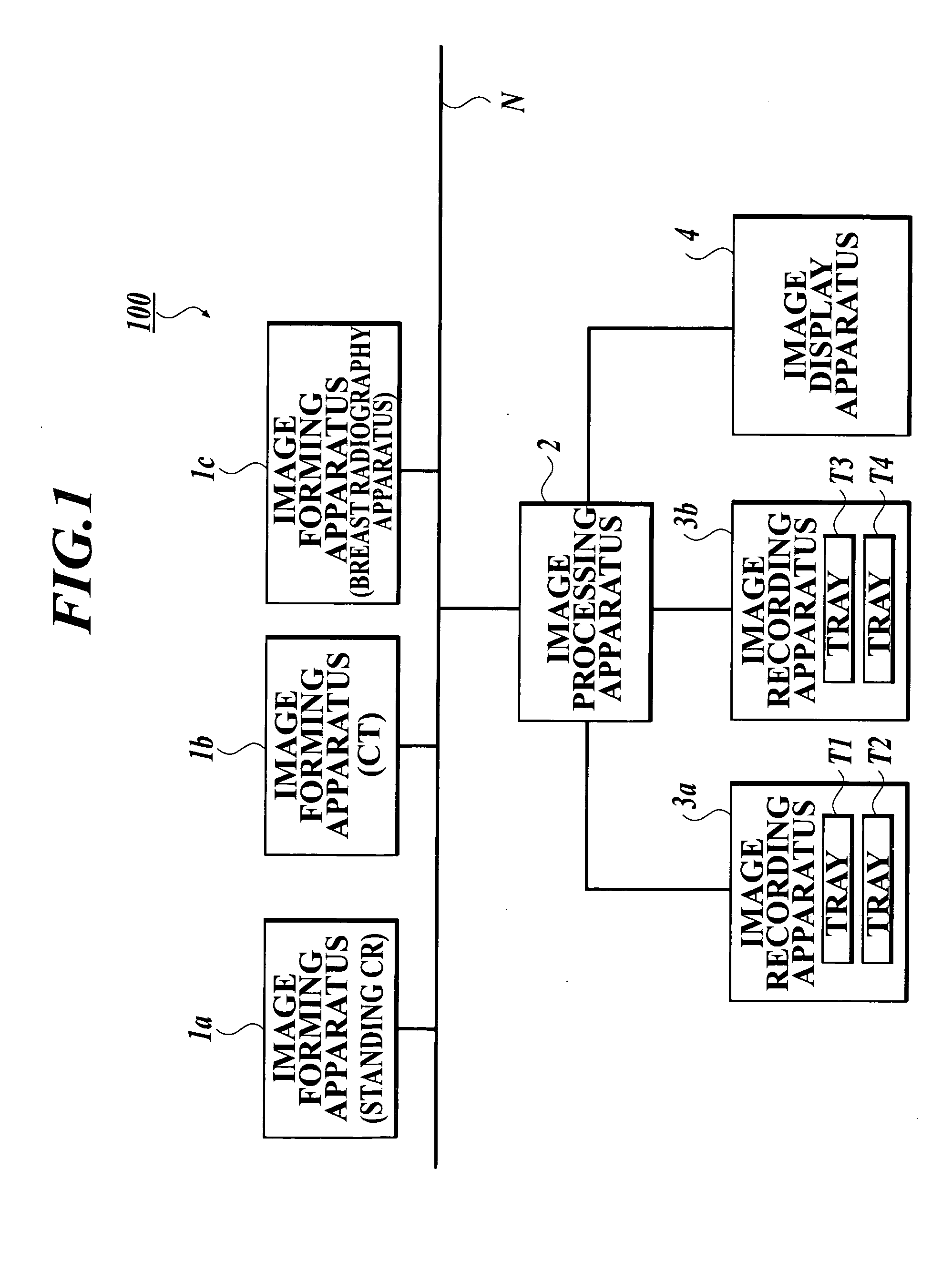
FIG. 1 is a conceptual diagram illustrating the entire configuration of the medical image processing system 100 in accordance with the embodiment. As illustrated in FIG. 1, the medical image processing system 100 is so connected that data can be transmitted and received between the image forming apparatus 1a-1c, and the image processing apparatus 2 through a network N. The image recording apparatus 3a, 3b, and the image display apparatus 4 are connected respectively to the image processing apparatus 2. The image processing apparatus 2 is so configured that data can be transmitted and received between the image recording apparatus 3a, 3b and the image display apparatus 4.

The network N can be various kinds of networks, being a LAN (Local Area Network), or a WAN (Wide Area Network), or an internet or the like. While wireless communication or infrared communication, when permitted by a medical in...


PUM

PropertyMeasurementUnit
sizeaaaaaaaaaa
pixel sizeaaaaaaaaaa
processing timeaaaaaaaaaa

Abstract

A medical image processing apparatus including: a diagnosis aid information generation section to generate diagnosis aid information by analyzing a medical image; a diagnosis aid information generation selection section to select whether to generate the diagnosis aid information, based on accessory information of the medical image; and an image output section to output the medical image and/or the generated diagnosis aid information to an image output apparatus.

Description

BACKGROUND OF THE INVENTION 1. Field of the Invention The present invention relates to a medical image processing apparatus which is capable of generating and outputting the corresponding diagnosis aid information by analyzing a medical image. 2. Description of Related Art A conventional system for aiding a doctor to diagnose is already known, wherein the diagnosis aid information such as a detection result of the abnormal shadow candidates is added to the medical image which is used for screening, and outputted to a CRT monitor, an LED, or the like. For example, a technique is disclosed which switches the image output destination corresponding to the detection result of the abnormal shadow candidates (e.g. refer to JP-Tokukai-2000-342559). The medical image and diagnosis aid information vary in output format and image output apparatus (e.g. a monitor, an image recording apparatus) with the individual diagnosis purpose as well as the image usage. However, the conventional appro...

Claims


Application Information

Patent Timeline
Patent Type & Authority Applications(United States)
IPC IPC(8): A61B5/00A61B6/00A61B6/03
CPCA61B6/00A61B6/461A61B6/566A61B6/4494G06T5/008G06T5/40G06F19/321G16H30/20G16H50/20
Inventor YANAGITA, AKIKO
Owner KONICA MINOLTA MEDICAL & GRAPHICS INC
Why Eureka
Try Eureka