
Method and system for automatic extraction of load-bearing regions of the cartilage and measurement of biomarkers

a technology of cartilage and load-bearing regions, which is applied in the field of automatic segmentation of the cartilage of the human knee, can solve the problems of incomplete information regarding the health of the cartilage over the whole of the cartilage, severe impact on the health of the knee joint, and inability to accurately assess so as to improve the diagnostic capability and the effect of better assessment of the health of the cartilag

Inactive Publication Date: 2005-05-26
VIRTUALSCOPICS
View PDF3 Cites 31 Cited by
  • Summary
  • Abstract
  • Description
  • Claims
  • Application Information

AI Technical Summary

Benefits of technology

[0013] Segmentation and the measurement of biomarkers, as techniques independent of each other, are known in the art. However, the inventors have discovered that the subdivision of cartilage into load bearing and non-load bearing regions provides a better assessment of the health of the cartilage, since in many conditions the load bearing region, which is more stressed, had earlier and more advanced changes in biomarker measurements. This examination of subregions thereby provides improved diagnostic capability over prior art which would measure biomarkers, such as cartilage volume or thickness, as a whole over the entire cartilage, thus combining information from both load bearing and non-load bearing regions of the cartilage.

Problems solved by technology

The knee joint can be severely affected by osteoarthritis (OA), which is the major cause of disabilities in older people.
Furthermore, knee injuries can create immediate major physical impairments via joint instabilities that will affect the joint load distribution or lead to the future development of OA.
This method uses the MRI data, an automated 3D segmentation, and an unsupervised joint space extraction algorithm that identify the medial and lateral compartments of the knee joint.
However, measurements over the whole of the cartilage do not provide complete information concerning the health of the cartilage.
The prior art provided no way to detect and assess those earlier and more advanced changes.
However, such techniques do not overcome the above-noted problems of the prior art.

Method used


Image

  • Method and system for automatic extraction of load-bearing regions of the cartilage and measurement of biomarkers
  • Method and system for automatic extraction of load-bearing regions of the cartilage and measurement of biomarkers
  • Method and system for automatic extraction of load-bearing regions of the cartilage and measurement of biomarkers

Examples


Embodiment Construction

[0023] A preferred embodiment of the present invention and experimental results therefrom will be set forth in detail with reference to the drawings, in which like reference numerals refer to like elements throughout.

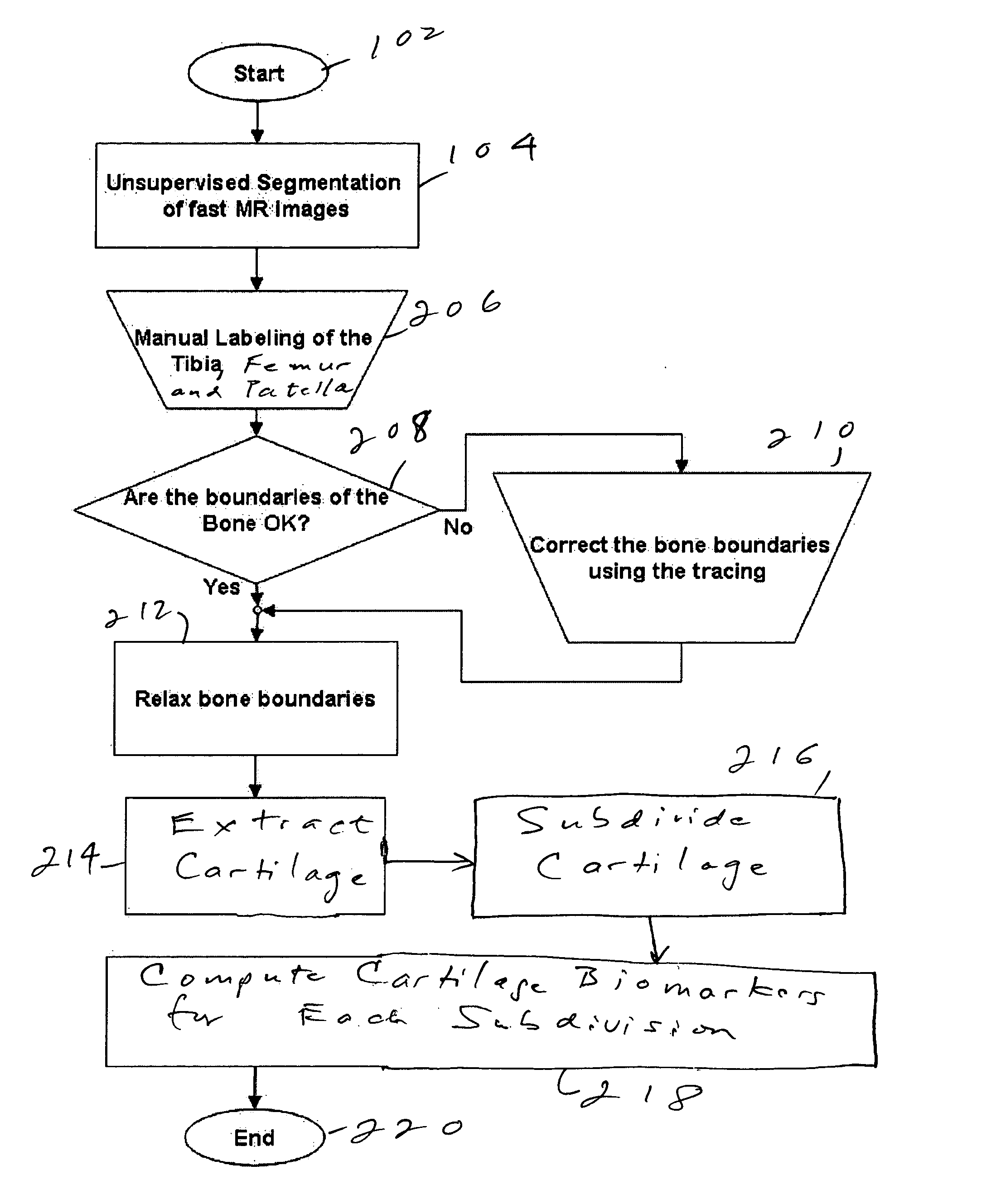
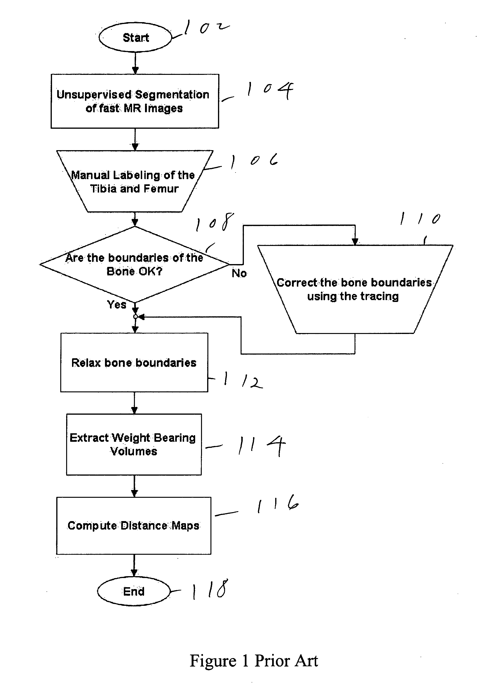
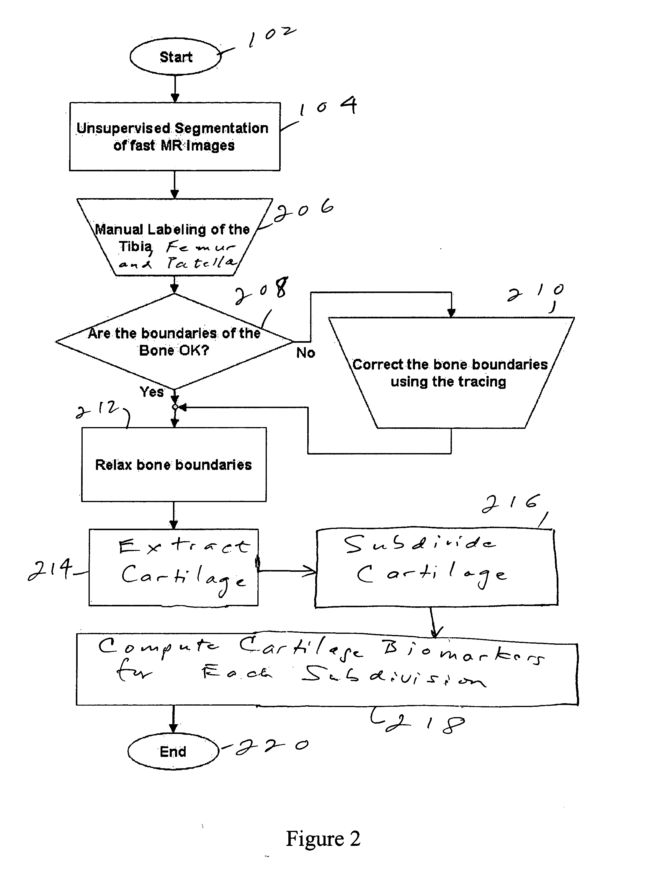
[0024]FIG. 2 shows a flow chart of the technique according to the preferred embodiment. Steps 102 and 104 are carried out like steps 102 and 104 of the prior technique of FIG. 1. However, in step 206, the tibia, femur, and patella are manually labeled. Steps 208, 210 and 212 are then carried out essentially like steps 108, 110 and 112 of FIG. 1, except that now the patella is also taken into account.

[0025] In step 214, the cartilage is extracted. In step 216, the cartilage is subdivided into subregions, in particular load-bearing and non-load-bearing subregions. In step 218, the cartilage biomarkers are computed for each subregion of the cartilage. The process ends in step 220.

[0026] We selected five MR image sets from three healthy adult subjects who had participate...


PUM


Abstract

An image is taken of a knee or other region of interest. The cartilage is extracted from the image and is subdivided into load-bearing and non-load-bearing regions. A biomarker is calculated for each of the load-bearing and non-load-bearing regions. The biomarkers can be assessed over time. The biomarkers for the load-bearing and non-load-bearing regions, and their changes, are used to assess the progress of joint disease.

Description

FIELD OF THE INVENTION [0001] The present invention is directed to a system and method for automatic segmentation of the cartilage of the human knee and more particularly to such automatic segmentation in which the cartilage is subdivided into a plurality of regions, including load-bearing regions and non-load-bearing regions. DESCRIPTION OF RELATED ART [0002] The knee joint can be severely affected by osteoarthritis (OA), which is the major cause of disabilities in older people. Furthermore, knee injuries can create immediate major physical impairments via joint instabilities that will affect the joint load distribution or lead to the future development of OA. [0003] In order to minimize the number of people with disabilities, the knee joint has been the focus of several studies that try to understand the knee mechanics and the nature of OA. The knee mechanics studies have focused on understanding the load distributions and the displacements of the knee under static or dynamic load...

Claims


Application Information

Patent Timeline
Patent Type & Authority Applications(United States)
IPC IPC(8): A61B5/055G06T5/00G06T7/00
CPCA61B5/055A61B5/103A61B5/4514G06T2207/30008G06T7/0012G06T7/0083G06T2207/10088A61B5/4528G06T7/12A61B5/704
Inventor TAMEZ-PENA, JOSETOTTERMAN, SAARA MARJATTA SOFIA
Owner VIRTUALSCOPICS
Learn More